Ileana Arsenescu
„Prof. Dr. C. C. Iliescu“ Emergency Institute for Cardiovascular Diseases, Bucharest
Dr. Ileana Arsenescu, “Prof. Dr. C. C. Iliescu” Emergency Institute for Cardiovascular Diseases, Bucharest
We present the Doppler curves recorded on the carotid and extracranial vertebral arteries, in a 72 year old patient, accusing a vertiginous syndrome
Cardiovascular risk factors: arterial hypertension, dyslipidemia, age.
Clinically: bilateral carotid murmurs can be detected.
Abreviations:
CCA – common carotid artery
ICA – internal carotid artery
ECA – external carotid artery
SV – systolic velocity
DV – diastolic velocity
RI – the resistive index
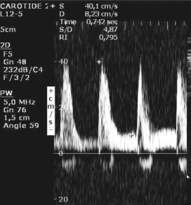
Figure 1. Right CCA; SV = 40 cm/a, DV = 8 cm/a, RI = 0.80 (Normal CCA RI ≤0.75). The velocities are below normal, especially the diastolic velocity, with an important RI increase.
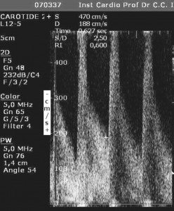
Figure 2. Right ICA (SV = 470 cm/s, DV = 188 cm/s, RI 0.60). Important increase of the systolic and diastolic velocities, meeting the criteria for a tight stenosis. Based on the velocity ratio ICA/CCA = 11.75 the stenosis is estimated at over 90%.
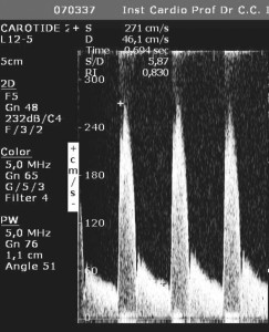
Figure 3. Right ECA (SV = 271 cm/s, VD = 46 cm/s, RI = 0.63). The velocity increase suggests a moderate stenosis located at the origin of the ECA. RI values remain elevated.
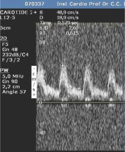
Figure 4. Right vertebral artery. SV = 49 cm/s, DV = 19 cm/s, RI = 0.61. The Doppler signal is normal.
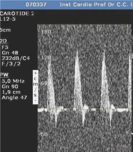
Figure 5. Right subclavian artery. Normal Doppler signal.
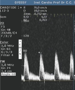
Figure 6. Left CCA. SV = 76 cm/s, DV = 18 cm/s, RI = 0.76 (Normal values for the CCA RI ≤0.75). The systolic velocity is normal, whereas the DV is slightly below normal values, pushing the RI a little over the normal values.
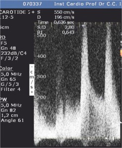
Figure 7. Left ICA. SV = 550 cm/s, DV = 196 cm/s, RI 0.64. Important increase of both systolic and diastolic velocities at the origin of the ICA, suggesting a tight stenosis at this level. ICA/CCA ratio is 7.2, compatible with a ≥80% stenosis.
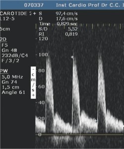
Figure 8. Left ECA. SV = 97 cm/s, DV = 18 cm/s, RI 0.82. The Doppler signal is normal.
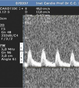
Figure 9. Left vertebral artery. SV = 48 cm/s, DV = 13 cm/s, RI = 0.73. (Normal RI <0.70). A mild increase of the RI, suggesting a possible distal stenosis.
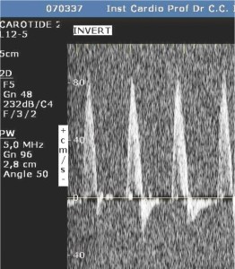
Figure 10. Left subclavian artery. Normal Doppler signal.
Comments
The patient had atheromatous plaques with important calcifications present on both internal carotid arteries, rendering an ulstrasonographic evaluation of the lesions impossible. Using Doppler criteria we were able to diagnose significant bilateral carotid stenosis.
The nominal values for velocity registered for the left ICA were higher than the ones in the right ICA. Thus, we might be tempted to conclude that the left carotid stenosis is more severe. However, the assessment of a stenosis should also consider the indirect criteria:
- Low systolic velocities were registered in the right CCA suggesting the presence of an obstacle on the lower trajectory of the artery, whereas the systolic velocities measured in the left CCA were normal
- The resistance index of the right CCA was significantly higher, another marker for the presence of a severe obstacle, while the one calculated for the left CCA was borderline normal, slightly over the superior limit
- The ICA/CCA ratio was higher on the right side, than on the left
Based on this data we can conclude that the most severe lesion is found on the right internal carotid artery, being estimated at more than 90%. In compensation, the blood flow in the left carotid axis increased, leading to higher velocities and an overestimation of the lesion when considering only the intrastenotic velocities.
Conflict of interests: none declared.
 This work is licensed under a