Dan Cristian Briscan1,2
1 University of Medicine and Pharmacy, Doctoral School of Biomedical Sciences, Oradea, Romania
2 Department of Internal Medicine II – Cardiology, Angiologyand Intensive Care Medicine, District Hospital Groß-Umstadt, Germany
Abstract: On patients with non-valvular atrial fibrillation, permanent anticoagulation at a score of CHA2DS2-VASc ≥1 is recommended. In relation to this, the risk of hemorrhage represented by HAS-BLED score increases. The concept of percutaneous interventional occlusion of the left atrial appendage is established more and more during the last years as an alternative to permanent therapy with anticoagulants on patients with non-valvular atrial fibrillation and with an incre-ased risk of hemorrhage. Imaging, through transesophageal echocardiograghy (TEE), has the main role in performing the percutaneous interventional occlusion of the left atrial appendage. Thus, pre-interventional, there should be established the patient’s selection, atria anatomy, left atrial appendage anatomy, procedures planning, optimum sizes for the device and the contraindications for the procedure should be excluded. Intra-procedural, in combination with fluoroscopy under angiographic control, the following are assured: transseptal puncture guidance, placing and anchoring the device, excluding residual flux, viewing intra-procedural complications such as, in our case, the formation of thrombotic material on the tip of Watchman device due to antithrombin-III deficiency and on this way, the procedural success of the intervention is being assured. Post-interventional, the complications assessment and occluder effi ciency on short and long term shall be assured. Keywords: atrial fibrillation – transesophageal echocardiograghy (TEE) – Watchman device – antithrombin-III defi ciency.
INTRODUCTION
On patients with atrial fibrillation without anticoagu-lants treatment, the global risk of stoke is 5 times hi-gher. Thus, this arrhythmia remains one of the major causes for stroke, sudden death and cardiovascular morbidity in the world. For this reason, permanent an-ticoagulation is recommended on patients with atrial fibrillation and a score of CHA2DS2-VASc ≥1 according to the current guide for atrial fi brillation treat-ment and management developed by the European Society of Cardiology (ESC)1. Drug therapy with non-vitamin K anticoagulants is very efficient in preventing the stroke and reduces the risk of brain hemorrha-ge in comparison with vitamin K antagonist therapy (VKA)2. The bleeding risk represented by HAS-BLED score is partially congruent with CHA2DS2-VASc score. According to the European Society of Cardi-ology (ESC), the relative contraindication regarding oral anticoagulants is represented for atrial fi brillation not only by HAS-BLED score, but also by the clinical assessment made by the attending physician, as well as by patient’s tendency of falling, for example, due to a posture and walking disorder3. Patients with con-traindication for permanent anticoagulation and with non-valvular fibrillation are represented in Garfi eld Registry (GARFIELD-AF Registry) by 3.2% of patients with CHA2DS2-VASc score between 0 and 1, as well as by 9.6 % of patients with a CHA2DS2-VASc score between 2 and 64.
Transesophageal echocardiograghy (TEE) is useful for additional assessment of valvulopathies and also for excluding intracardiac thrombi, especially on left atrial appendage (LAA) in order to facilitate early cardioversion or catheter ablation5. Once it was ob-served that approximately 90% of thrombi have been produced on non-valvular atrial fi brillation on the left atrial appendage (LAA), systems for its occlusion and closure have been developed by means of interventi-onal occlusion6-9. One of these devices is represented by the Watchman device (Boston Scientific), which represents one of the best devices scientifically studi-ed. Through transesophageal echocardiograghy (TEE), more measurements of the left atrial appendage (LAA) are performed, on at least 4 angles of echocardiogra-phic views which are: at 0, 45, 90 and 135°. Through that, one can precisely determine the opening of the left atrial appendage (LAA) respectively the size of the Watchman device. Additionally 3D transesophageal echocardiograghy can be used in order to quantify the opening surface of the left atrial appendage (LAA). The anatomical morphologies of the left atrial appendage (LAA) most commonly encountered are: the windso-ck, the broccoli, the cactus and the chicken wing.
From these different morphologies, the chicken wing is the most frequent one10. In case of a com-plex anatomy of the left atrial appendage (LAA), pre-interventional measurements can be made through computerized tomography (CT) or through magnetic resonance tomography (MRT).
Watchman device implantation technique
On most facilities, this procedure takes about 30-45 minutes and is minimally invasive. This procedure is performed under sedation; general anesthesia is not required. Before beginning the procedure, intracardi-ac thrombi and pericardial effusion are excluded and optimum sizes of the device are determined in accor-dance with the left atrial appendage (LAA) anatomy using transesophageal echocardiograghy (TEE). After the device implantation, this should present a com-pression of about 72% up to 90% from its nominal di-ameter, according to the manufacturer´s instructions.
After the right femoral vein puncture and after placing the catheter into the right atrium, transsep-tal puncture is performed at the proper height of the septum, posterior on the inferior septal area, under fluoroscopic guidance by means of Pigtail catheter, respectively echographic control by means of intra-procedural transesophageal echocardiograghy (TEE) (Figure A – image 1 and 2).
Through the transseptal guide wire, the guiding ca-theter with a thickness of 12-14 French is being in-troduced for the occlusion system. Once the air from the occlusion system is being eliminated by means of saline NaCl injection, its inclusion into the transsep-tal guiding catheter follows. Under fl uoroscopic and echographic control, the device is being placed and an-chored into the left atrial appendage (LAA). After that, Tug test is being performed in order to verify optimal placement and stability of the Watchman device. In case of a suboptimal placement of the device, this will be retraced into the occluder system and then, its op-timal placement will follow. After its fi nal placement, a contrast agent fluoroscopic representation respecti-vely a test for the presence of residual flux are perfor-med. This will be done on the side part of the device through transesophageal echocardiograghy (TEE) as well as through 3D transesophageal echocardiogra-ghy. Once the Watchman device is correctly placed, its release from the screw connection of the occluder system will be the next step. On the day following the device implantation, the presence of pericardial effusi-on must be excluded.
This procedure is made under the control of acti-vated coagulation time (ACT), anticoagulation being performed with unfractionated heparin (UFH).
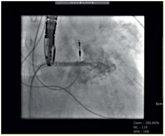
Figure A. Interventional occlusion of the left atrial appendage by means of a 27 mm Watchman device guided by angiographic control and intra-procedural TEE. Image 1. After transseptal puncture the Watchman device is guided inside the left atrial appendage (LAA), highlighting its anatomy through fluoroscopy after administrating the contrast agent by means of the Pigtail catheter.
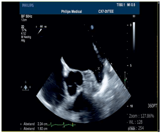
Image 2. Establishing optimal sizes of the device depending on the anat-omy of the left atrial appendage (LAA) through transesophageal echocar-diograghy (TEE) on an echocardiographic view angle of 44°.
CASE PRESENTATION
The 84 years old patient came in for elective hospita-lization, respectively for the left atrial appendage in-terventional occlusion with a Watchman device due to bleeding complications occurred under new anti-coagulants treatment. According to the patient’s me-dical history the following are known: transient ische-mic attack (TIA) with transitory aphasia, symptomatic epilepsy with complex focal seizures, decompensated heart failure with pleural fluid on the left side, medium tricuspid insufficiency, mild aortic insufficiency, mild mitral insufficiency, atrial fibrillation with a 7 points CHA2DS2-VASc score and a risk for stroke of 10% per year, a 5 points HAS-BLED score and thus ha-ving a very high hemorrhagic risk, cardiovascular risk – hypertension (HT), repeated upper digestive hemor-rhage with iron deficiency under anticoagulant therapy with rivaroxaban within GAVE syndrome (gastric an-tral vascular ectasia) with folate deficiency.
On admission, the patient’s health condition was generally good: from the cardiovascular point of view – stable; with normal clinical, local and neurological examination, without significant information. Medica-tion: Bisoprolol 2.5 mg, Ramipril 2.5 mg, Torasemi-de 10 mg, Nitrendipine 5 mg, Levetiracetam 250 mg, Oxycodone 30 mg, Omeprazole 20 mg, Folsäure 5 mg, Ferro Sanol 50 mg, Calcium D osteo, Lactulose 10 ml.
Pre-interventional transesophageal echocardiogra-ghy (TEE) was ambulatory performed into the cardio-logy office of the medical care centre.
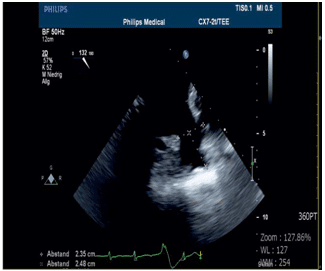
Image 3. Respectively on an echocardiographic view angle of 132°.
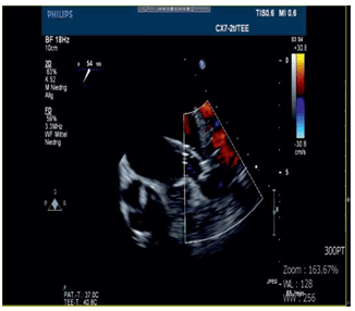
Image 4. Device placement and anchorage into the left atrial appendage (LAA).
Interventional occlusion of the left atrial appendage
During the intervention, once the right femoral punc-ture respectively the transseptal puncture were per-formed without any complications, the guiding cathe-ter and Watchman device were introduced. Throm-botic material was formed on the tip of the Watchman device viewed on transesophageal echocardiograghy.
Then, the device was replaced by a multifunctional 8 F catheter in order to suck out over and over again the biggest parts of the thrombotic material. Because of some small residual parts of the thrombotic material on the guiding catheter, the decision to stop the inter-vention was made. Thus, an 8 F catheter has been re-introduced and, under continuous suction the guiding catheter from the sept is being withdrawn. That way, all residual parts of the thrombotic material could be eliminated. When the control of activated clotting time (ACT) was made, only therapeutic values were determined, the anticoagulation being made with un-fractionated heparin (UFH).
During laboratory examination in hemostasis, an-tithrombin-III defi ciency was found. This caused the thrombotic material formation during the interven-tion. Post-interventional, no other complication was found, both imaging and clinically.
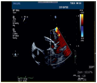
Image 5. Tug test in order to verify optimal placement and stability of the Watchman device.
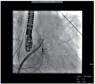
Image 6. Fluoroscopy performance. The Watchman device nitinol struc-ture is being revealed in occlusion position, after its release from the screw connection of the occlusion system.
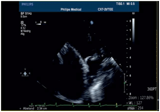
Image 7. Echocardiographic control by means of device size measurement after implant at a 0° view angle.
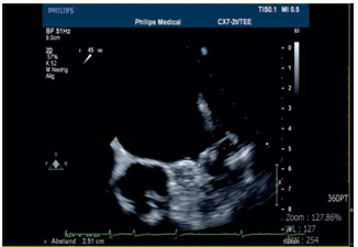
Image 8. Respectively a 45° view angle.
Antithrombin (antithrombin-III)
Antithrombin represents the main inhibitor of blood clotting. This mainly inactivates factors Xa, IXa and IIa (thrombin) enzymes ans also performs inactivation ac-tions over factors XIIa, XIa, VIIa complex and tissue factor. In adults, concentrations below 40-70% are associated with a tendency to thrombosis11. Congeni-tal antithrombin deficiency is very rare. The reduction of acquired antithrombin concentration is to be diffe-rentially considered depending on clinic signs, therefo-re, this is less significant on hepatic diseases than on consumptive coagulopathy12. Causes for antithrombin deficiency can be represented: by synthesis disorders (very rarely congenital, liver diseases, asparaginase treatment), in increased consumption (consumptive coagulopathy – disseminated intravascular coagulation (DIC)), in inflammatory processes (systemic inflamma-tory response syndrome, sepsis, systemic hyperfibri-nolysis), in loses (nephrotic syndrome, ascites, wounds with large surface, massive blood transfusions) or on newborn babies (physiological).
After 4 weeks, a 27 mm Watchman device has been implanted under antithrombin-III substitution, witho-ut any complication.Therapy with Clopidogrel 75 mg has been recommended for 6 months and Aspirin 100 mg for the rest of her life. A control should be made through transesophageal echocardiograghy after a pe-riod of 3 and 6 months from the intervention.
Transesophageal echocardiograghy (TEE)
The role of imaging in percutaneous interventional occlusion of the left atrial appendage is extremely es-sential and irreplaceable, having a pre-interventional, intra-procedural and post-interventional role.
By using pre-interventional transesophageal echo-cardiograghy we can establish patient´s selection, atria anatomy, fossa ovalis identification, left atrial appen-dage anatomy, procedures planning, optimum sizes of the device and we can exclude the contraindications in performing this procedure. Contraindications are represented by the pre-existence of: an interatrial shunt, atrial septal aneurysm, intracardiac thrombi or left atrial appendage thrombi, pericardial effusion, mi-tral stenosis with orifice area smaller than 1.5 cm2, a cardiac masses or tumours, complex aterom with mobile plate in the aortic arch or descendent aorta, as well as by placing the device, interference with any intracardiac or intravascular structure.
By using intra-procedural transesophageal echocar-diograghy in combination with fl uoroscopy we can as-sure the guidance of septal puncture, device placement and anchorage and we can exclude residual flux. In our case, we could visualize the formation of thrombotic material on Watchman device due to antithrombin deficiency and this way we could immediately and totally remove the thrombotic material without any other interventional complications.
By means of post-interventional transesophageal echocardiograghy we can assure complications assess-ment, such as exclusion of an iatrogenic shunt in the interatrial septum, of pericardial effusion, of residual flux or the appearance of a new paraprotetic flux or the appearance of thrombi on the device. We can also assess the effi ciency, stability and the position of the occluder on short and long term.
CONCLUSION
The Watchman device, through endotelialisation, per-manently eliminates the possibility of thrombi forma-tion on the left atrial appendage (LAA) and so, the risk of stroke on non-valvular atrial fi brillation is being considerably reduced. It represents an alternative to anticoagulant treatment. Anticoagulant treatment in-terruption on long term, after performing interventio-nal occlusion of the left atrial appendage, can eliminate or visible reduce the hemorrhagic risk. This way, the Watchman device can represent a cheaper alterna-tive, as an unique procedure that eliminates the risk of a stroke, from which a specific group of patients could benefi t. As a result, percutaneous interventio-nal occlusion of the left atrial appendage is the only procedure that could considerably reduce hemorr-hagic risk on patients with triple therapy (antiplatelet double therapy and anticoagulant). The success rate of this procedure rises above 95%, depending on the left atrial appendage (LAA) anatomy as well as on the experience of the team that performs the procedure.
Echocardiography represents imaging examination as main role, through which percutaneous interven-tional occlusion of the left atrial appendage is perfor-med. That way, the thickness of the device could be determined based on the left atrial appendage (LAA) anatomy, intra-procedural complications could be vi-ewed in real time – as in our case, thrombotic material formation on Watchman device due to antithrombin-III defi ciency – and major complications could be avoid.
Conflict of interest: none declared.
References
1. Kirchhof P, Benussi S, Kotecha D et al (2016) 2016, ESC Guidelines for the management of atrial fi brillation developed in collaboration with EACTS. Eur Heart J37:2893–2962.
2. Ruff CT, Giugliano RP, Braunwald E et al (2014).Comparison of the efficacy and safety of new oral anticoagulants with warfarin in pa-tients with atrial fibrillation: a meta-analysis of randomised trials. Lancet383:955–962.
3. O’Brien EC, Holmes DN, Ansell JE, Allen LA, Hylek E, Kowey PR, Gersh BJ, Fonarow GC, Koller CR, Ezekowitz MD, Mahaffey KW, Chang P, Peterson ED, Piccini JP, Singer DE (2014). Physician prac-tices regarding contraindications to oral anticoagulation in atrial fi-brillation: fi ndings from the Outcomes Registry for Better Informed Treatment of Atrial Fibrillation (ORBIT-AF) registry. AmHeart J167:601–609.e1.
4. Kakkar AK,Mueller I, Bassand JP, FitzmauriceDA, Goldhaber SZ, Goto S, Haas S, Hacke W, Lip GY, Mantovani LG, TurpieAG, van EickelsM, Misselwitz F, Rushton Smith S, Kayani G, Wilkinson P, Ver-heugt FW, Registry Investigators GARFIELD (2013). Risk profiles and antithrombotic treatment of patients newly diagnosed with atrial fi brillation at risk of stroke: perspectives from the international, ob-servational, prospective GARFIELD registry. PLOSONE8:e63479.
5. Conti A, Canuti E, Mariannini Y, Viviani G, Poggioni C, Boni V, Pini R, Vanni S,Padeletti L, Gensini GF. Clinical management of atrial fi-brillation: early interventions, observation, and structured follow-up reduce hospitalizations. Am J EmergMed 2012;30:1962–1969.
6. Bajaj NS, Parashar A, Agarwal S, Sodhi N, Poddar KL, Garg A, Tuz-cu EM,Kapadia SR. Percutaneous left atrial appendage occlusion for stroke prophylaxis in nonvalvular atrial fibrillation: a systematic re-view and analysis of observational studies. JACC Cardiovasc Interv 2014;7:296–304.
7. Lewalter T, Kanagaratnam P, Schmidt B, Rosenqvist M, Nielsen-Kudsk JE,Ibrahim R, Albers BA, Camm AJ. Ischaemic stroke preven-tion in patients with atrial fibrillation and high bleeding risk: oppor-tunities and challenges for percutaneous left atrial appendage occlu-sion. Europace 2014;16:626–630.
8. Meier B, Blaauw Y, Khattab AA, Lewalter T, Sievert H, Tondo C, Glikson M.EHRA/EAPCI expert consensus statement on catheter-based left atrial appendage occlusion. Europace 2014;16:1397–1416.
9. Holmes DR Jr, Kar S, Price MJ, Whisenant B, Sievert H, Doshi SK, Huber K,Reddy VY. Prospective randomized evaluation of the Watch man Left Atrial AppendageClosure device in patients with atrial fibrillation versus long-term warfarin therapy: the PREVAIL tri-al. J Am Coll Cardiol 2014;64:1–12.
10. Di Biase L, Santangeli P, Anselmino M, et al. Does the left atrial ap-pendage morphology correlate with the risk of stroke in patients with atrial fibrillation? Results from a multicenter study. J Am Coll Cardiol. 2012;60(6):531–8.
11. Hirsh J (1977) Hypercoagulability. Semin Hemat 14:409–425.
12. Lechner K, Kyrle PA (1995) Antithrombin III concentrates – are they clinically useful? Thromb Haemost 73(3):340–348. 5. Yano Y et al. Isolated Systolic Hypertension in Young and Middle-Aged Adults, and 31-Year Risk for Cardiovascular Mortality: the Chicago Heart Association Detection Project in Industry Study. J.ACC 2015; 65: 327-335.
 This work is licensed under a