Download PDF
https://doi.org/10.47803/rjc.2020.30.3.443
Mihai Melnic1, Flavius Gherasie1, Al Hassan Ali1, Ionut Stanca1, Mihai Santa1, Cristian Girgel1, Adrian Linte1, Serban Balanescu1,2
1 Elias University Emergency Hospital, Bucharest, Romania
2 University of Medicine and Pharmacy „Carol Davila”, Bucharest, Romania
Coronary artery disease (CAD) is a pathological pro-cess characterized by atherosclerotic plaque accumulation in the epicardial arteries, obstructive or non-obstructive. The disease can be stable but can also become unstable at any time, typically due to an acute atherothrombotic event caused by plaque rupture or erosion. New-onset angina is generally regarded as unstable angina. Emergency angiographic evaluation is recommended.
Left main coronary artery disease has a higher prognostic risk as a result of the large myocardial territory at risk, ranging from 75% to 100%, depending on the dominance of the left coronary circulation.
Current clinical practice guidelines from both the American College of Cardiology/American Heart Association and the European Society of Cardiology recommend revascularization for all patients with ≥50% stenosis of the left main coronary artery (LM), regardless of symptomatic status or associated ischemic burden.
Drug-eluting stents are preferred for LM revascu-larization as they offer improved survival and fewer adverse cardiovascular events compared with BMS.
In a study of 57 de novo left main bifurcation (LMB) lesions, TAP stenting with second-generation DES had acceptable TLF rate at 3-year follow-up of 13.3%. In contrast the BBK (Bifurcations Bad Krozingen) II study, where 39% had LM stenting, the Culotte technique compared to TAP had at one year, a TLF of 6.7% vs 12.0% (P = 0.11).
Current practice guidelines in support of revascularization of all LM lesions ≥ 50% are based on older trials in an era when medical therapy was limited and before the use of invasive physiological assessment of stenosis severity. In fact, the same evidence suggests that medical management of patients at lower risk might be associated with favorable outcomes. Although smaller studies support the use of FFR and IVUS to define lower-risk groups with LM disease who could be treated by optimal medical therapy alone, larger trials assessing clinical outcomes over longer follow-up are needed to fully assess this strategy.
MEDICAL MANAGEMENT OF LMCAD
In the COURAGE (Clinical Outcomes Utilizing Revascularization and Aggressive Drug Evaluation) trial2, similar outcomes were observed for an initial strategy of optimal medical therapy compared with initial revascularization in patients with stable non-LM CAD. The safety of deferred revascularization in patients with stable LM disease is less well known, but current clinical practice guidelines strongly recommend revas-cularization in all patients with ≥50% stenosis of the LM. The basis for these class IA recommendations is predicated on post hoc data derived from subgroup analyses of 185 patients with LM disease from 2 randomized clinical trials (RCTs) of patients with chronic stable angina conducted in the late 1970s and early 1980s3,4 demonstrating the superiority of surgical revascularization over medical therapy on 5- to 10-year survival. These early RCTs were conducted in an era when medical therapy was, by contemporary standards, limited. For example, only 66% of „medically managed” patients with LM in those early trials received -blockers and only 19% were taking aspirin. These trials antedated the current widespread use of disease-modifying pharmacologic interventions (such as statins, inhibitors of the renin-angiotensin-aldosterone system, and more effective antiplatelet agents such as P2Y12 inhibitors), which reduce adverse car-diovascular events in patients with CAD.
Current clinical guidelines strongly recommend surgical revascularization for LMCAD (class IA) with PCI considered a reasonable alternative (class II) in select patients with less complex anatomy (SYNTAX score of <33) and clinical characteristics that predict an increased risk of adverse surgical outcomes. How-ever, patients with PCI need close clinical follow-up, as they may have a higher need for repeat revascularization in the future.
CASE PRESENTATION
A 31 years old male patient with cardiovascular risk factor (mild dyslipidemia) without any prior presentation for cardiovascular disease, is admitted on the Cardiology Department with a diagnosis of de novo angina (unstable angina). The patient describes typical retrosternal pain with radiation on both arms. The current symptoms appeared 2 months ago. The patient also did a a positive ECG stress test (Bruce protocol) on the day of the presentation at Elias Hospital. The general objective examination reveals a stable general condition, normal rhythmic heartbeats without any heart murmurs and no pulmonary or sistemic congestion. The electrocardiogram (Figure 1) performed on admission shows no particular ischemic modifications and the cardiac enzymes value levels are in the normal range. Complete blood count and bio-chemistry laboratory tests are within normal range, with mild dysiidemia (LDL=77 mg/dl, HDL=29mg/dl, TG=163mg/dl). Echocardiography shows a nondilated left ventricle (LV) with preserved systolic function (ejection fraction 60%), with no wall motion abnormalities, no significant valvulopathies, and no signs of pulmonary hypertension.
The patient is transported to the cathlab for emergency coronarography. The coronarography shows the right coronary artery (RCA) without significant stenosis, subocclusion of left main, subocclusion of ostial left anterior descending artery (LAD), 70% steno-sis of the mid segment and subocclusion of the circumflex artery –Medina 1-1-1 bifurcation lesion (Figure 2, 2a the arrows point at the stenosis).
Single stent technique, with or without final kissing balloon inflation, should be preferred over the 2-stent technique, whenever possible, to minimize stent length or stent overlap. The multiple 2-stent techniques need to be utilized, however, when tackling compromised or dissected large side branches. We describe a provisional 2-stent technique, TAP technique, which has the characteristic of minimizing stent overlap at the bifurcation and ensuring good stent coverage at the side branch ostium.
The NORDIC I trial compared a provisional versus planned 2-stent technique with drug-eluting stents. There was only a 2.7% crossover rate to the 2-stent technique in the provisional group, and no difference in MACE between the two groups at 6 months. A 6 Fr lumen EBU 3.5 guiding catheter was chosen, radial approach. Predilatation was performed in the LAD, LCX and left main arteries. A 3.0 x 24 mm DES (Biomatrix alpha – Biosensors) was delivered into the mid left anterior descending artery (LAD) (Figure 3,4), Then a 4.0 x 24 mm DES (Biomatrix alpha – Biosensors) was deployed at high pressure from the left main into the LAD (Figure 5,6), however, the result was suboptimal in the ostial circumflex artery, we manage to re-cross into the circumflex artery.
We performed predilation with a 2.0 x 15mm semicompliant ballon infl ated throught stent struts into the ostium of the circumflex artery. We delivered a 3.5 x 14 mm DES (Biomatrix alpha – Biosensors) that was pulled back 1-2 mm into the left main and inflated at 18 atm (Figure 7,8).
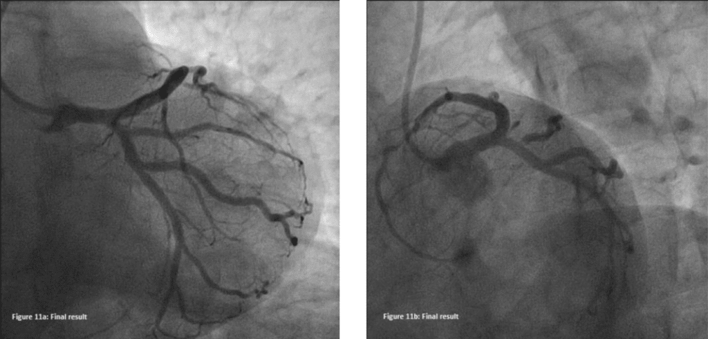
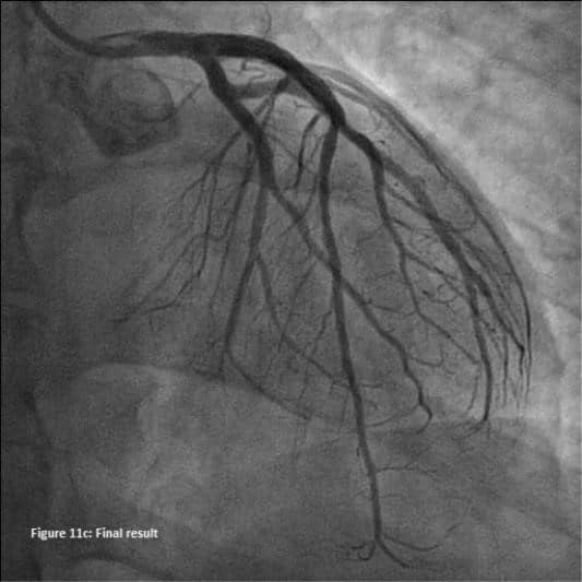
Multiple kissing inflations were performed in the distal left main bifurcation (Figure 9), and finally, POT was performed with a 5.0 x 8 mm noncompliant balloon inflated at 12-14 atm pressures throughout the left main (Figure 10,11a, 11b,11c).
The patient remains anginafree at 4 months post intervention.
Patient was put on medical therapy consisting of ticagrelor 90 mg twice/day, AAS 75 mg once/day, ator-vastatin 80 mg once/day, bisoprolol 5 mg once/ day and pantoprazole 40 mg once/day.
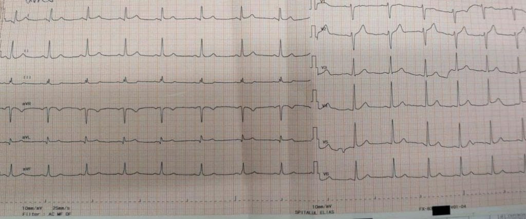
Figure 1. ECG at presentation showing sinus rhythm at 75 beats, no particular ischemic modifications.
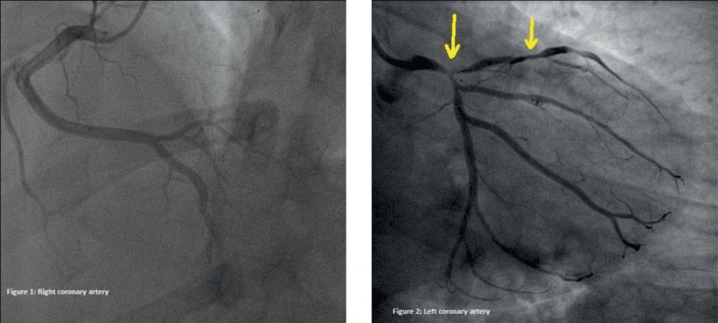
Figure 2. Figure 2a.

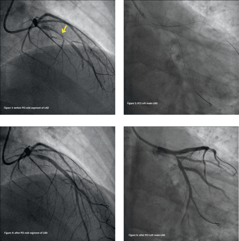
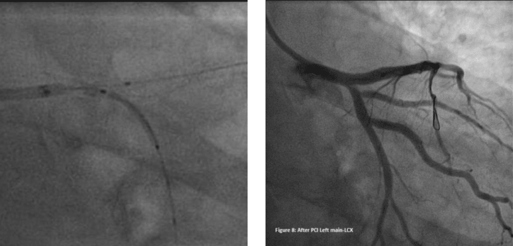
Figure 7. TAP technique.
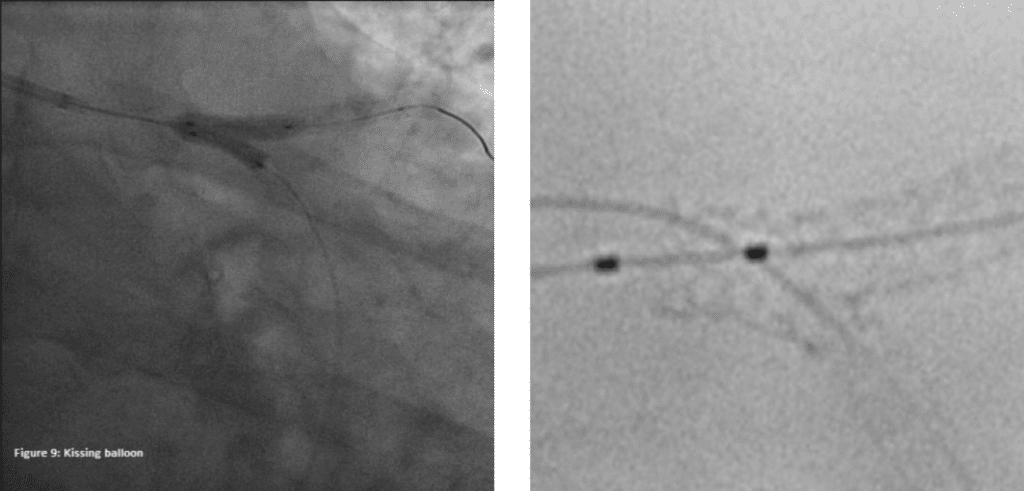
Figure 7. StentBoost® after final POT.
DISCUSSION
The modification of the T-stenting was first described in 2007 by Burzotta et al1. It was evaluated in vitro and in two independent series of patients undergoing elective drug-eluting stent (DES) implantation on a bifurcation lesion. In vitro testing demonstrated perfect coverage of the bifurcation with minimal stent’s struts overlap at the proximal segment of SB ostium with a single layer stent struts. Sirolimus, paclitaxel, or zotarolimus DES were deployed in 73 patients (67% with Medina 1,1,1 lesions and 44% of unprotected distal left main disease) using the TAP technique. The procedural success was achieved in all cases. At 9 months the clinically-driven target vessel revascularization (TVR) was 6.8%. Since this was a pilot study, the investigators recommended larger outcome trials to further evaluate this technique. It is expected that the results of EXCEL and NOBLE will determine the next guidelines for the foreseeable future, as forthcoming trials of this magnitude are unlikely to be pursued from economic and priority viewpoints unless marked advances in revascularization technologies emerge. Importantly, a heart team approach for shared decision-making should be the standard of care for all cases of LMCAD.
CONCLUSION
In comparison with T-stenting, the TAP technique with kissing balloon inflation and final POT is associated with a reduced rate of restenosis in the side branch.
The TAP technique is a provisional 2-stent technique that involves stenting the main branch, and in the presence of a suboptimal ostial side-branch result, passing a second stent into the side branch with 1–2 protruding into the main, followed of kissing inflation si final POT optimisation.5-12
The benefit of this „TAP” technique (1-2mm over-lap), is that the opportunity to first perform single-stenting is preserved, while side-branch stenting can be performed only if required. This technique would also minimize the overlap of drug-eluting stents, similar to the mini-crush technique, thereby potentially reducing the risk of stent thrombosis.13-21 Recrossing into the side branch in both cases becomes reasonably simple, since with this technique, only one layer of stent needs to be crossed at any given time.
Conflict of interest: none declared.
References
1. Park SJ, Kim YH, Park DW, Lee SW, Kim WJ, Suh J, Yun SC, Lee CW, Hong MK, Lee JH, Park SW. Impact of intravascular ultrasound guidance on long-term mortality in stenting for unprotected left main coronary artery stenosis. Circ Cardiovasc Interv. 2009; 2:167– 177.
2. Buszman PE, Buszman PP, Kiesz RS, Bochenek A, Trela B, Konkol-ewska M, Wallace-Bradley D, Wilczynski M, Banasiewicz-Szkrobka I, Peszek-Przybyla E, Krol M, Kondys M, Milewski K, Wiernek S, Debinski M, Zurakowski A, Martin JL, Tendera M. Early and long-term results of unprotected left main coronary artery stenting: the LE MANS (Left Main Coronary Artery Stenting) registry. J Am Coll Cardiol. 2009; 54:1500–1511.
3. Morice MC, Serruys PW, Kappetein AP, Feldman TE, Stahle E, Co-lombo A, Mack MJ, Holmes DR, Choi JW, Ruzyllo W, Religa G, Huang J, Roy K, Dawkins KD, Mohr F. Five-year outcomes in pa-tients with left main disease treated with either percutaneous coro-nary intervention or coronary artery bypass grafting in the synergy between percutaneous coronary intervention with taxus and cardiac surgery trial. Circulation. 2014; 129:2388–2394.
4. Stone GW, Sabik JF, Serruys PW, Simonton CA, Genereux P, Pus-kas J, Kandzari DE, Morice MC, Lembo N, Brown WM, Taggart DP, Banning A, Merkely B, Horkay F, Boonstra PW, van Boven AJ, Ungi I, Bogats G, Mansour S, Noiseux N, Sabate M, Pomar J, Hick-ey M, Gershlick A, Buszman P, Bochenek A, Schampaert E, Page P, Dressler O, Kosmidou I, Mehran R, Pocock SJ, Kappetein AP. Evero-limus-eluting stents or bypass surgery for left main coronary artery disease. N Engl J Med. 2016; 375:2223–2235.
5. Levine GN, Bates ER, Blankenship JC, Bailey SR, Bittl JA, Cercek B, Chambers CE, Ellis SG, Guyton RA, Hollenberg SM, Khot UN, Lange RA, Mauri L, Mehran R, Moussa ID, Mukherjee D, Nallamothu BK, Ting HH. 2011 ACCF/AHA/SCAI guideline for percutaneous cor-onary intervention: executive summary: a report of the American College of Cardiology Foundation/American Heart Association Task Force on Practice Guidelines and the Society for Cardiovascular An-giography and Interventions. Circulation. 2011; 124:2574–2609
6. Veterans Administration Coronary Artery Bypass Surgery Coopera-tive Study Group. Eleven-year survival in the Veterans Administra-tion randomized trial of coronary bypass surgery for stable angina. N Engl J Med. 1984; 311:1333–1339.
7. Varnauskas E. Twelve-year follow-up of survival in the randomized European Coronary Surgery Study. N Engl J Med. 1988; 319:332– 337.
8. Conley MJ, Ely RL, Kisslo J, Lee KL, McNeer JF, Rosati RA. The prog-nostic spectrum of left main stenosis. Circulation. 1978; 57:947–952.
9. Murphy ML, Hultgren HN, Detre K, Thomsen J, Takaro T. Treat-ment of chronic stable angina. A preliminary report of survival data of the randomized Veterans Administration cooperative study. N Engl J Med. 1977; 297:621–627.
10. Takaro T, Peduzzi P, Detre KM, Hultgren HN, Murphy ML, van der Bel-Kahn J, Thomsen J, Meadows WR. Survival in subgroups of pa-tients with left main coronary artery disease. Veterans Administration Cooperative Study of Surgery for Coronary Arterial Occlusive Disease. Circulation. 1982; 66:14–22.
11. Prospective randomised study of coronary artery bypass surgery in stable angina pectoris. Second interim report by the European Cor-onary Surgery Study Group. Lancet. 1980; 2:491–495.
12. Prospective randomised study of coronary artery bypass surgery in stable angina pectoris. Second interim report by the European Cor-onary Surgery Study Group. Lancet. 1980; 2:491–495.
13. MRC/BHF Heart Protection Study of cholesterol lowering with sim-vastatin in 20,536 high-risk individuals: a randomised placebo-con-trolled trial. Lancet. 2002; 360:7–22.
14. Colombo A, Stankovic G, Orlic D, et al. Modified T-stenting with crushing for bifurcation lesions: Immediate results and 30-day out-comes. Catheter Cardiovasc Interv 2003;60:145–151.
15. Ge L, Airoldi F, Iakovou I, et al. Clinical and angiographic outcome after implantation of drug-eluting stents in bifurcation lesions with the crush stent technique: Importance of final kissing balloon post-dilation. J Am Coll Cardiol 2005;46:613–620.
16. Ge L, Iakovou I, Cosgrave J, et al. Treatment of bifurcation lesions with two stents: One-year angiographic and clinical follow up of crush versus T-stenting. Heart 2006;92:371–376.
17. Hoye A, Iakovou I, Ge L, et al. Long-term outcomes after stenting of bifurcation lesions with the “crush” technique: Predictors of an adverse outcome. J Am Coll Cardiol 2006;47:1949–1958.
18. Galassi AR, Colombo A, Buchbinder M, et al. Long-term outcomes of bifurcation lesions after implantation of drug-eluting stents with the “mini-crush technique.” Catheter Cardiovasc Interv 2007;69:976– 983.
19. Moussa I, Costa RA, Leon MB, et al. A prospective registry to evalu-ate sirolimus-eluting stents implanted at coronary bifurcation lesions using the “Crush technique.” Am J Cardiol 2006;97:1317–1321.
20. Burzotta F, Gwon HC, Hahn JY, Romagnoli E, Choi JH, Trani C, Colombo A. Modified T-stenting with intentional protrusion of the side-branch stent within the main vessel stent to ensure ostial cov-erage and facilitate final kissing balloon: the T-stenting and small pro-trusion technique (TAP-stenting). Report of bench testing and first clinical Italian-Korean two-centre experience. Catheter Cardiovasc Interv.2007;70:75-82.
21. Al Rashdan I, Amin H. Carina modification T stenting, a new bifurca-tion stenting technique: clinical and angiographic data from the first 156 consecutive patients. Catheter Cardiovasc Interv. 2009;74:683-90.22. Dzavik V, Kharbanda R, Ivanov J, et al. Predictors of long-term outcome after crush stenting of coronary bifurcation lesions: Impor-tance of the bifurcation angle. Am Heart J 2006;152:762–769.
22. Jim MH, Wu EB, Fung RC, Ng AK, Yiu KH, Siu CW, Ho HH. Angio-graphic result of T-stenting with small protrusion using drug-eluting stents in the management of ischemic side branch: the ARTEMIS study. Heart Vessels.2014 Mar 14.
23. Collins N, Dzavik V. A modified balloon crush approach improves side branch access and side branch stent apposition during crush stenting of coronary bifurcation lesions. Catheter Cardiovasc Interv 2006;68:365–371.
24. Brunel P, Lefevre T, Darremont O, Louvard Y. Provisional T-stent-ing and kissing balloon in the treatment of coronary bifurcation le-sions: Results of the French multicenter “TULIPE” study. Catheter Cardiovasc Interv 2006;68:67–73.
25. Burzotta F, Dzavik V, Ferenc M, Trani C, Stankovic G. Technical as-pects of the T And small Protrusion (TAP) technique. EuroInterven-tion 2015;11 Suppl V:V91-5.
26. Jabbour RJ, Tanaka A, Pagnesi M et al. T-Stenting With Small Protru-sion: The Default Strategy for Bailout Provisional Stenting? J Am Coll Cardiol Interv 2016;9:1853-4.
27. Ferenc M, Gick M, Comberg T et al. Culotte stenting vs. TAP stent-ing for treatment of de-novo coronary bifurcation lesions with the need for side-branch stenting: the Bifurcations Bad Krozingen (BBK) II angiographic trial. Eur Heart J 2016. https://www.ahajournals.org/doi/10.1161/JAHA.117.008151
 This work is licensed under a