Serban Balanescu1
1 Department of Cardiology, Elias Emergency University Hospital, „Carol Davila” University of Medicine and Pharmacy, Bucharest, Romania
Abstract: Takotsubo phenotype is defined as an acute, reversible heart failure syndrome precipitated by an acute stress-ful event occurring mainly in old post-menopausal women. Typical clinical presentation is a form of acute coronary syndro-me with apical ballooning. Currently the condition is considered as a syndrome with multiple causes, all associated with acute stress and excess catecholamine release from the pituitary-adrenal axis. Generalized use of coronary angiography lead to increased recognition of this syndrome, as far as a majority of patients has normal epicardial arteries or minor coronary atherosclerosis. Recent data sustain inclusion of Takotsubo syndrome with acute coronary syndromes in between ST-ele-vation and non ST-elevation ACS.
Although initially regarded as a reversible cause of myocardial dysfunction with complete recovery and good outcome, re-cently multiple follow-up studies demonstrated that prognosis in Takotsubo syndrome is similar to that of acute coronary syndromes. As far as Takotsubo may arise from extremely different clinical conditions, a recent position paper proposed a sub classifi cation of this condition depending on the offending event. This is sustained by different outcome of Takotsubo related to etiology.
The current paper reviews recent data about causes, mechanisms, outcome and classifi cation of Takotsubo syndrome.
Keywords: Takotsubo syndrome, acute coronary syndrome, catecholamine excess, neuro-endocrine imbalance.
INTRODUCTION AND DEFINITION: A NECESSARY REAPPRAISAL
Ever since its first description by Sato et al in the early ‘90s in a Japanese conational1, Takotsubo syndrome (TS) was viewed as a puzzling Cardiology dilemma despite extensive basic and clinical research2. The idiom “between the devil and the deep blue sea”, used to express how difficult it is to choose between two undesirable situations reflects the enigmatic appearan-ce and vanishing of this disease. Due to its striking wall motion dyssynergy, mimicking a severe acute co-ronary syndrome and surprisingly normal angiographic coronary appearance the disease was named initially “tako-tsubo-like left ventricular dysfunction” by the Japanese group who first identified it1,3.
The disease occurs mainly in post-menopausal women, aged more than 55 years in their sixth to seventh decade, precipitated mainly by acute negative or positive mental or physical stress. The typical cli-nical presentation is that of suspected acute coronary syndrome with either ST-elevation or ST-depression associated with acute systolic left ventricular dysfunction4. While both ST segment changes and wall motion abnormalities may be striking on admission, they are generally transient and resolve in 6 weeks; they may variably persist up-to 3-to-6 months from the index event5.
TS differs significantly from acute myocardial in-farction, because not only is there no coronary artery stenosis, but segmental dyskinesia extends beyond the distribution territory of a single coronary artery6. The presentation mimicking an acute MI with normal epicardial coronary arteries raised the suspicion of an acute form of cardiomyopathy, vigorously denied by some researchers7. Up-to-date Takotsubo pathophy-siology hypotheses suggest the disease may be viewed as a mix between and abnormal sympathetic cardi-omyocyte response and microvascular dysfunction6.
Mental or physical stress may be recognized as a precipitating factor in more than 80% of Takotsubo patients8. The disease was called „stress cardiomyopathy”, „broken heart syndrome”, „happy heart syndrome” and „voodoo heart” to emphasize the im-portance of psychological or psychiatric factors in the pathophysiology of this clinical syndrome.
Currently Takotsubo is considered a syndrome that may be induced by a large variety of precipitating factors that have in common sympathetic overstimu-lation of the myocardium and catecholamine-induced myocardial stunning, myocardial injury and reversible myocardial dysfunction5,6.
EPIDEMIOLOGY
Today TS occurs in about 2% of patients with an initial diagnosis of ACS, but can reach 5.9-7.5% of ACS in women9.
There is a major increase in the reported incidence of TS in the last 5-10 years. Data reported from diffe-rent world regions and health systems demonstrate more and more cases of TS. In the SCAAR Registry of all-comers with an acute coronary syndrome with or without an acute heart failure syndrome TS incidence increased more than 10 times, from 0.16% in 2005 to 2.2% in 201210. In the USA between 2007 and 2012 from a total of 53.947 patients with a Takotsubo dis-charge diagnosis the prevalence increased from 52 in 2007 to 178 per million discharges in 201211.
The increased prevalence may be due to a real in-crease in cases due to progressively higher psychosoci-al stress of modern life or to increased awareness and widespread use of coronary angiography in suspected acute coronary syndromes10. The constant increase of cases allowed better description of causes, pathophy-siological correlates and to establish treatment effect and clinical outcomes12.
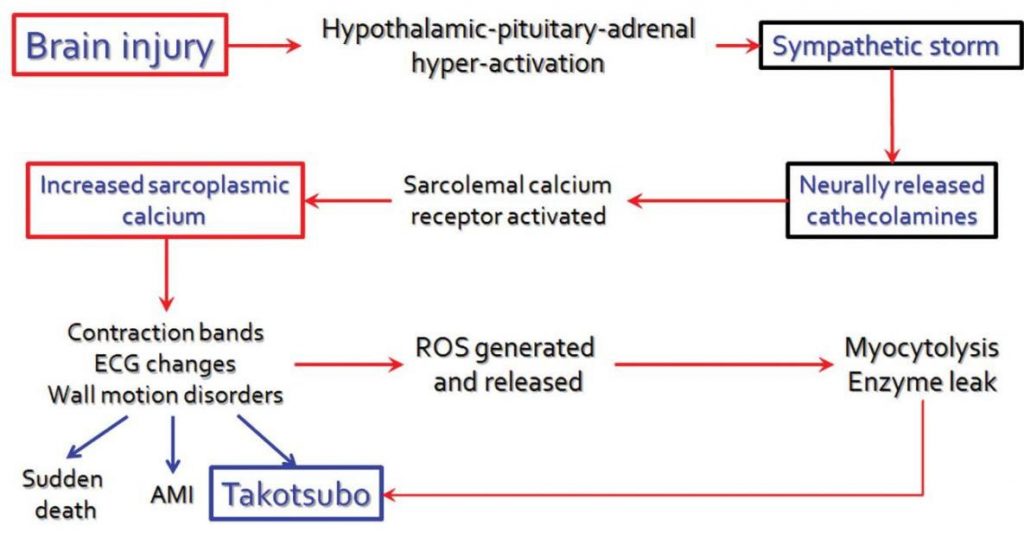
Figure 1. The proposed mechanism of adrenergic-mediated myocardial injury in TS due to central activation of the sympathetic system.
ETIOLOGY
The InterTAK Registry is an international database consisting of a consortium of 26 worldwide centers initiated in Zurich that currently follows-up more than 2000 TS patients all-over the world4. Most of the data regarding TS are obtained from this registry13,14.
The recently published International Expert Con-sensus on TS enlists the extremely numerous and varied physical and emotional triggers of the disease (Table 1)6. As recognized from the initial cases, psychi-atric disease has major impact on triggering TS. Mood disorders were diagnosed in 39% of TS cases, anxiety disorders in 27%, while major personality disorders such as schizophrenia occurs in a minority of 1% of Takotsubo patients15. Anxiety, negative affectivity and obsessive-compulsive personality are more frequently observed, while isolated depression or neuroticism is seldom found16. Resting state functional brain MRI in TS patients demonstrated specific areas with increased connectivity located in the precuneus region as opposed to healthy controls16. Other functional MRI studies demonstrated activation of the hippocampus, the brainstem, basal ganglia and prefrontal cortex in acute TS patients that subsided at 30-day follow-up17. All these brain regions are involved in stimulation of the locus coeruleus as the main structure responsible for sympathetic hyper activation.
Another frequently observed patient subset presen-ting with TS are individuals with acute neurological di-sease. „Neurogenic myocardial injury” associated with myocardial stunning is a condition determined by ne-urologic events of different significance18. In a national US registry of hospitalized patients admitted between 2006 and 2014 comprising 9.146.013 cases with neu-rologic disease, 5832 patients were discharged with a TS diagnosis19. Any acute neurological condition on admission had an OR of developing TS of 1.54 (95% CI: 1.48 to 1.62); the highest risk of developing TS was found in status epilepticus (OR=4.9; 95% CI:3.7-6.3) and subarachnoid hemorrhage (OR=11.7; CI 95%: 10.2-13.4)19. However all other acute neurologic di-sease presented an increased risk for developing TS, starting with ischemic stroke, intracerebral hemorrha-ge, migraine or meningoencephalitis. Recently TS was recognized even in patients with coronary artery disease (CAD) (Figure 2). As far as TS is associated with any cause of acute psychic or so-matic stress and acute myocardial infarction is a major stress factor associated with increased serum cathe-colamines, TS and CAD may occur together in the same patient. TS may be diagnosed in a CAD patient if the severely diseased coronary artery responsible for an ACS is distributed to a totally different myocar-dial territory than the one showing the wall motion anomalies20. Some cases are reported with STEMI due to occlusion of a diagonal vessel and a widely patent LAD, with severe apical ballooning and hyperkinetic basal segments21.
Another supporting argument for the association between TS and CAD is the patient population that can typically develop TS: old post-menopausal women (Figure 3). Advanced age and associated risk factors for CAD may explain concomitant occurrence of TS and coronary atherosclerosis. In an intracoronary imaging study in 23 patients with TS that were investigated by optical coherence tomography (OCT) in the left main and LAD during an ACS, 69.6% of subjects presented atherosclerotic plaques while 26.1% had thin-cap fi-broatheromas22. No thrombus or properly ulcerated coronary plaques were found by OCT, emphasizing that CAD is an incidental finding in TS.
Various other clinical situations were described in association with TS such as accidental administration of large subcutaneous adrenaline injection23 or after treatment with check-point inhibitors for different ne-oplasia24.
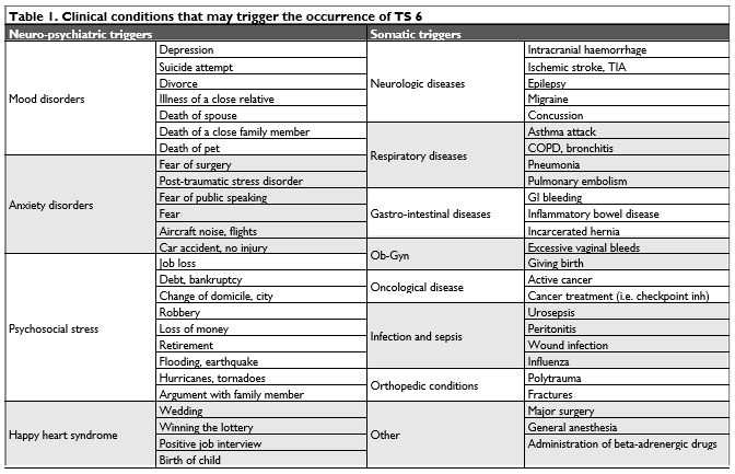
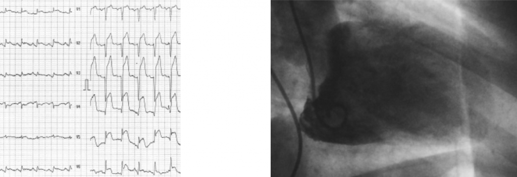
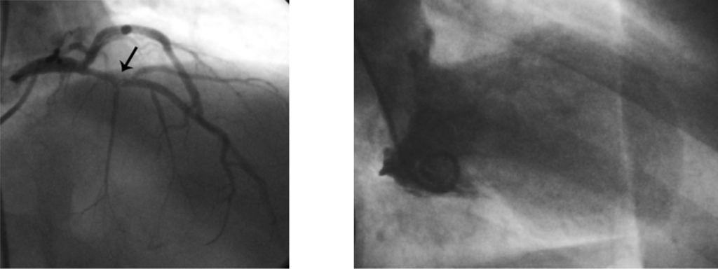
Figure 2. A Takotsubo patient with noncritical coronary artery disease. The 58 year-old patient was admitted because of acute STEMI with significant ST elevation in V2-V6 and infero-lateral leads (Figure 2A). Coronary angiography showed a moderate proximal LAD stenosis involving the first major septal branch (black arrow in Figure 2B). LV angiogram (in diastole (Figure 2C) and in systole (Figure 2D) showed a large apical ballooning syndrome involving the inferior wall of LV and hyperkinetic basal segments.
PATHOPHYSIOLOGY
There are four main hypotheses that try to explain the occurrence of TS25,26:
Myocardial impairment due to excessive local catecholamine availability by abnormal epinephri-ne/norepinephrine release via both hypothala-mic-pituitary-adrenal axis and local release from
sympathetic nerve endings. Patients with TS have a 2-to-3 fold increase of serum cathecolamines versus patients with STEMI27,28. The catecholami-ne excess mediates myocardial injury by direct toxicity, epicardial and microcirculatory vaso-constriction and increased cardiac afterload2.
Beta-2 adrenoceptor / Gi-dependent cardio de-pression. Activation of the beta-2 receptor by epinephrine. Activation of inhibitory G protein. Apical dysfunction due to apex-to-base gradient of beta-2 receptors.
Focal perfusion defects and microvascular ob-struction or coronary spasm. TS was recently included with the acute coronary syndromes due to microvascular dysfunction29. Presenting with either ST elevation or depression, positive biomarkers for myocardial injury (higher NT-pro-BNP and lower troponin than in ACS) and typically normal angiographic appearance of co-ronary arteries, TS is currently considered in the spectrum of ACS together with STEMI, Prinzme-tal angina and NSTEMI29.
Acute, focal metabolic down-regulation in res-ponse to demand / supply mismatches similar to ischemic stunning. Form of protective reaction against malignant arrhythmias and/or necrosis.
The typical neurogenic-mediated myocardial injury is observed in acute stroke. The insular cortex anato-mically located in the Sylvian sulcus is responsible for complex functional integrates, both motor and senso-ry in correlation with the limbic system18. It controls autonomic nervous system maintaining the balance between sympathetic and vagal systems30. Ischemic lesions of the insula were proven to increase serum catecholamine levels and induce myocardial dysfuncti-on31. Interestingly stimulation of the right insula leads to sympathetic hyperactivity, while stimulation of the left insula leads to parasympathetic dominance32. Insular cortex is usually involved with the parietal lobe in stroke due to middle cerebral artery occlusion33.
The main site of noradrenergic production in the brain is the locus coeruleus that concentrate all sti-muli from hypothalamus, amygdala and the neocortex, including the insula and the frontal cortex, triggering the systemic sympathetic response34. Norepinephri-ne production in the locus coeruleus activates the hypothalamic-pituitary-adrenal axis8.
Myocardial cathecolamines are a combination between locally released norepinephrine from the sympathetic nerve endings and the serum epinephrine
– norepinephrine released by the adrenal medulla35. In normal conditions cathecolamines stimulate beta-1 adrenergic receptors and increase intracellular cAMP intermediated by a second-messenger mechanism through the Gs protein family. Catecholamine excess may switch cellular adrenergic effects from beta-1 re-ceptor stimulation to beta-2 receptor activation cou-pled with an inhibitory second-messenger Gi prote-in36. Beta-2 receptors are distributed mainly toward the apex: the apico-basal gradient of these receptors is considered responsible for the apical ballooning syndrome, but it does not explain the other middle-segment or basal forms of TS37. Calcium sarcoplasmic overload induced by beta-adrenoceptor stimulation results in contractile dysfunction by ATP depletion38. Direct negative effects of cathecolamines on the car-diomyocytes are completed by adrenergic-mediated activation of inflammation and oxidative stress39 (Fi-gure 1).
Neurogenic myocardial injury induces different effects on cardiomyocytes than myocardial ischemia and necrosis. Neurogenic injury produces myofibrillar degeneration and myocytolysis distributed surroun-ding the epicardial nerve fibers, while myocardial ne-crosis occurs mainly in the subendocardial layers and respects distribution of a coronary artery40.
It has been postulated that apical ballooning and catecholamine activation of beta-2 receptors and the increased activity of inhibitory G protein may repre-sent a mechanism for cardiomyocyte protection to limit myocardial damage of sudden marked increase in epinephrine41. Myocardial dysfunction occurs in the apical region where there is the largest concentration of epinephrine sensitive beta-2 receptors, while the basal regions displaying the largest concentration of beta-1, norepinephrine sensitive receptors remain unaffected42. These findings suggest that TS phenotype may be epinephrine specific43.
Microvascular catecholamine-mediated vasospasm may contribute to pathophysiology of TS39. Observed perfusion defects in the acute setting are completely reversible by adenosine injection and at follow-up study44.
Thus myocardial stunning and systolic dysfunction in TS may represent a protective mechanism against a metabolism/flow mismatch: increased catecholamine normally lead to increased metabolic demands, while microvascular dysfunction reduces supply of oxygen, glucose and free fatty acids. Myocardial stunning wo-uld protect cells from self-destruction and would allow complete subsequent recovery when catechola-mine levels return to normal.

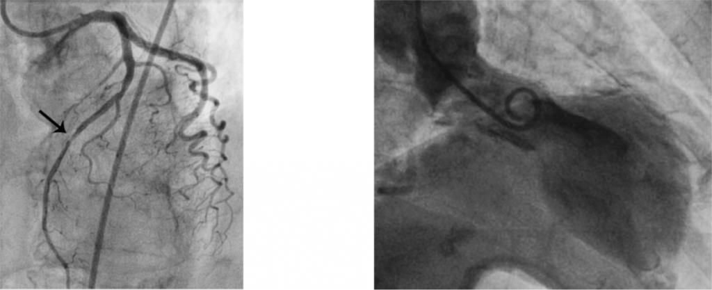
Figure 3. A 67 year-old depressive woman with an acute stressful event presents with chest pain, signs and symptoms of LV failure and ST-elevation in the first 4h from symptom onset (Figure 3A). Coronary angiography demonstrates a moderate mid-LAD stenosis (black arrow in Figure 3A). Takotsubo syndrome is diagnosed after LV angiogram shows typical apical LV ballooning (Figures 3C and 3D). Coexisting Takotsubo and non-signifi cant obstructive CAD may be identified in old patients with CV risk factors.
CLINICAL PRESENTATION
Patients with TS present usually with symptoms, signs and ECG patterns of an ACS45. These may occur in a patient with an acute neurological event. Sometimes the neurological event is diagnosed after angiography shows no coronary artery stenosis in a comma patient with cardiac arrest, making differential diagnosis diffi – cult between the causative factors: neurologic disea-se associated with TS or cardiac arrest with hypoxic neurologic damage. However, prolonged chest pain and dyspnea are the most common symptomatic pat-terns of TS onset, followed by acute LV failure and pulmonary edema. Syncope, cardiac arrest or cardio-genic shock are also encountered in TS, although not frequently.
When compared with age- and sex-matched pati-ents with acute coronary syndromes (ACS), TS pati-ents have less chest pain, more severe systolic LV dys-function and significantly less obstructive coronary ar-tery disease 4. There is no difference in the prevalence of cardiogenic shock (10-12% of patients) or 30-days mortality (4-5%) between TS and ACS counterparts4. Neurologic (either acute or chronic) or psychiatric disorders are strikingly more encountered in TS pati-ents. Acute neurologic disease is found 9 times more frequently and psychiatric disorders are found three times more in TS patients.
Major complications of TS replicate all common complications of ACS9. Mechanical complications ex-tend from overt LV failure and pulmonary edema (12-45% of cases), acute mitral regurgitation (14-25%) or cardiogenic shock (6-20%) to free wall or interven-tricular septum rupture (less than 1%)5. A particular complication of TS is acute LVOT obstruction as far as antero-basal segments are hyperkinetic and the dyski-netic apex pulls the anterior mitral leafl et towards the septum in the LVOT46. Apical thrombus formation and systemic embolization were described in 2-8% of ca-ses. QT prolongation, brady and tachyarrhythmia with sudden cardiac death were also mentioned in 2-5% of patients. Paroxysmal atrial fi brillation occurs in 5 to 15% of TS cases5.
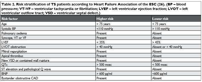
A plethora of clinical, echocardiographic and serum biological markers were identified to correlate with high risk in TS patients on admission (Table 2)26.
ECHOCARDIOGRAPHY AND ANGIOGRAPHY FINDINGS
Starting from the initial description of TS as an “apical ballooning syndrome”47, four different patterns of seg-mental wall motion anomalies are accepted today as parts of the spectrum of the disease4,14.
- The apical, typical pattern with apical ballooning, mimicking antero-apical LV aneurysm is the most frequently encountered in 81.7% of cases.
- The mid-ventricular pattern is associated with hyperkinetic apical and basal segments while middle segments of the anterior and inferior wall are dyskinetic. This pattern is observed in 14.6% of cases.
- The basal type is characterized by apical hyperki-nesia while the basal segments are dyskinetic. It is found in 2.2% of cases.
- The focal type is associated with focal dyskinesia of any myocardial segment, i.e. the mid segment of the anterior wall, while all other segments are contracting normally. It is the most seldom
found, in 1.5% of cases.
Occasionally a Takotsubo-like wall motion dyski-nesia with cardiogenic shock, fully reversible, was de-scribed to involve the free wall of the right ventricle without any signs of acute pulmonary hypertension or embolism48, but this TS presentation was not included with the typical cases yet.
Apparently, the type of acute stressor event may produce different patterns or phenotypes of TS. Ne-gative stressors induce more frequently apical ballooning in the “broken heart syndrome”, while positive stressors or “happy heart” may be associated with mid-ventricular TS49.
Sometimes it is very difficult to differentiate in clini-cal practice between ACS, myocarditis and TS, mainly when coronary angiography identifies no significant obstructive coronary artery disease. Contrast magnetic resonance imaging should be used to clarify this issue, as apparently there are specific patterns in each and every of these conditions50.
DIAGNOSIS AND DIFFERENTIATION FROM ACS
Issued in 2008 the modified Mayo diagnostic criteria were used to differentiate TS from other acute heart failure syndromes47. The following diagnostic criteria were considered:
Transient hypokinesis, akinesia or dyskinesia of mid-LV segments with or without apical involve-ment. Wall segment anomalies extend beyond myocardial distribution of a single coronary vessel. Previous stress factors are frequently found.
Absence of obstructive coronary disease or angi-ographic evidence of acute plaque rupture. New ECG changes (either ST-elevation and/or T-wave inversion) or modest elevation in cardiac troponin.
Absence of pheochromocytoma or myocarditis. The Mayo criteria were reviewed and modified by the Heart Failure Association of the ESC in 2016 who added recovery of ventricular function on cardiac ima-ging at 3-6 months follow-up as a supplemental crite-rion26. The data obtained from the InterTAK registry lead to the recently published International Takotsubo Diagnostic Criteria issued by the ESC in 20186.
Because TS has a significant presentation overlap with acute coronary syndromes and acute heart fa-ilure, accurate diagnosis is mandatory in order to prescribe appropriate investigations and treatment. Data from the InterTAK Registry allowed develop-ment of a clinical score (“InterTAK diagnostic score”) that should ease differentiation between TS and acute coronary syndromes51 (Table 3). There are 7 major criteria included, female sex and emotional trigger be-ing the most significant. When a cut-off value of 70 points is considered, there is a high probability (more than 90%) of TS over 70 points and a low-intermediate probability of TS with a score of less than 70 points5.
When compared with ACS, TS patients have lower troponin and higher BNP serum levels; the extent of ventricular motion anomalies greatly exceeds necrosis biomarker increase, reflecting the stunned, recovera-ble myocardium5.
Some research teams demonstrated that circulating micro-RNAs (19-25 nucleotides) that are non-coding transcriptional regulators of complex cellular functi-ons (such as apoptosis, proliferation or differentiation) could be used to distinguish between TS and acute MI52. Eight micro-RNAs were selected for verification by real-time quantitative reverse transcription PCR. In this trial patients with TS had a significant up-regulati-on of miR-16, miR-26a and let-7F, while patients with STEMI had miR-1 and miR-133a up regulated with res-pect to TS. Interestingly the identified micro-RNAs are up regulated by stress and depression confirming the close relationship between TS and neuropsychia-tric disease.
Consequently establishing the diagnosis of TS evol-ved from excluding major coronary disease or myo-carditis to specific criteria53.

TREATMENT OF TS
There are no prospective randomized trials in TS pa-tients. Thus all recommendations are based on clinical experience and have a class C (expert consensus) le-vel of evidence. Current data based on retrospecti-ve studies, meta-analysis and small case series were gathered in a recently published International Expert Consensus Document5.
There is general consensus to admit all patients with TS in the Coronary Care Unit where prompt full clinical, ECG, echocardiographic, angiographic and serum biomarkers should allow classification of risk (Table 2)26.
Lower risk patients with LVEF higher than 45% need no specific treatment and should be closely followed-up until recovery. Low-risk patients with LVEF betwe-en 35-45% should be treated with progressively in-creasing doses of beta-blockers and ACE-inhibitors. Early discharge strategy may be considered in these low-risk category patients. Complex cardiac imaging by echocardiography and cardiac MRI should be per-formed at follow-up to confirm myocardial recovery and exclude myocardial infarction or myocarditis26.
High-risk patients should be kept in CCU for at least 72h. Beta-blockers should be carefully initiated although LVEF may be lower than 45%; patients with ventricular tachycardia or fibrillation and those with paroxysmal atrial fibrillation also benefit from beta-blockers. LVOT obstruction with a maximum gradient more than 40 mmHg is another indication for beta-blockers. ACE-inhibitors should be given in all patients with LVEF lower than 45%. Apical thrombus needs low-molecular weight heparin (LMWH) acutely and subsequent oral anticoagulation for at least 3 months. High-risk patients should be treated however with a LMWH for 7 days, even when no intraventricular thrombosis can be found. Acute pulmonary edema should be treated with standard doses of IV loop diu-retics and nitroglycerine.
Cardiogenic shock is the most difficult clinical pre-sentation with TS as systemic cathecolamines are spontaneously high, are implicated in the pathoge-nesis of the disease and may worsen myocardial dys-function when administered as therapeutic measure to increase LV systolic function. TS patients treated with cathecolamines have a 20% mortality rate4. An ECMO machine and/or LVAD or Impella device may be necessary to properly support heart function and systemic oxygenation until LV function recovers. Le-vosimendan as a calcium-sensitizer, non-beta-recep-tor-dependent positive inotropic drug may be empi-rically used to increase LV systolic function54. Shock TS patients with LVOT obstruction cannot be treated with beta-blockers and may benefi t from ivabradine as bradycardia-inducing drug in sinus rhythm55.
Long-term treatment comprises ACE-inhibitors or sartans, while no benefit of chronic beta-blocker use could be demonstrated even on the recurrence of TS4,56. If CAD is present at angiography, but it cannot be held responsible for clinical presentation while the patient is diagnosed with TS, appropriate doses of a statin and aspirin should be started and continued in-definitely.
There are no data regarding the effect of treating anxiety, mood disorders or any associated psychiatric disease on the occurrence of TS, which is another to-pic for controversy.
OUTCOME
TS is far from being a benign condition, despite the fact it is regarded as an acute reversible heart failu-re syndrome. Initial reports considered the disease as benign, based on the rapid recovery of left ventricular function when compared with ACS57.
Cardiogenic shock occurs in TS with the same pre-valence as in atherothrombotic ACS4. Cardiogenic shock in TS was observed at admission in 11.4% of 711 patients enrolled in the RETAKO Registry58. In-hospital mortality of TS is 4-5% similar with that of STEMI reperfused by primary PCI59,60. 30-day morta-lity is 5.9% and the long-term mortality rate is 5.6%/ patient/year4.
Long-term outcome of TS patients is similar to tho-se with CAD and worse than matched control sub-jects as demonstrated in a group of 505 patients from the SCAAR Registry60. TS patients have a 2.1 times higher risk for mortality at 5 years when compared to control subjects without CAD, despite having low cardiovascular risk profile.
Despite prompt (sometimes spontaneous) reco-very of left ventricular function long-term outcome in TS may not be as benign as previously thought. In a trial on 37 patients with previous TS more than 1 year from acute presentation they had impaired car-diac deformation indices and lower cardiac energetic markers61. Thus it is currently considered that TS pati-ents may develop a persistent subclinical heart failure phenotype.
Recently data from the InterTAK Registry show-ed that the long-term outcome of TS depends on the initial trigger12. Patients with a pure emotional trigger and no other major comorbidity have the best 5-year outcome, while TS patients due to neurologic disor-ders have the highest 5-year mortality that may reach 20%12. Based on this difference in outcome depending on the triggering factor, the InterTAK Registry inves-tigators recently proposed a new classification of TS. Class I is considered TS due to emotional triggers. Class II consists of TS patients due to physical stress; class IIa is due to medical conditions or interventions and intense physical activities; class IIb is due to neu-rologic disease and has the worst outcome. Class III is TS without any identifiable cause. Time will tell if this classification confirms its usefulness in TS patients.
Data obtained from 371 patients with TS enrolled in an Italian registry and followed-up for a mean of 26+/-20 months demonstrated that CHA2DS2-VASc score could be used to predict death, MI or stroke in a similar way with cardioembolic risk prediction in patients with atrial fibrillation62. The patients were di-vided in three groups with respect to the calculated CHA2DS2-VASc score on enrollment: 0-1, 2-3 and hi-gher than 4. Patients with CHA2DS2-VASc score ≥ 4 had a MACCE prevalence of 17% while those with a CHA2DS2-VASc score £ 1 had a MACCE prevalence of 6% (p=0.033).
The recurrence rate of TS is a topic of controversy; it is considered TS may recur in up-to 22% of all TS patients45. The triggering factor and TS phenotype may be different from the those seen with the first TS occurrence63.
CONCLUSION
Takostubo syndrome is an acute reversible heart fa-ilure syndrome with various segmental wall motion anomalies, mostly with apical ballooning according to the originally described, typical pattern. Some form of acute stress is always elicited in personal history and there is a complex interaction with neuro-psychiatric disorders and increased plasma cathecolamines.
TS may be associated with coronary disease com-monly non obstructive and stable. In some cases Takotsubo may occur associated with an acute coro-nary syndrome (as a major stressful event), with wall motion anomalies extending beyond coronary distri-bution of the responsible artery.
Although generally reversible between 2 to 6 months after the index event, TS has the same com-plex prognosis and complications as all other acute coronary syndromes. The worst recognized outcome occurs when TS is associated with any acute neuro-logic disease. Risk stratifi cation should identify higher risk patients (i.e. cardiogenic shock or severe acute left ventricular failure), needing intensive treatment and supervision. Standard treatment of acute heart failure is recommended, while avoiding beta-mimetic drugs in this condition associated anyway with serum catecholamine excess.
Financial support: none declared.
Conflicts of interest: none declared.
References
1. Dote K, Sato H, Tateishi H, Uchida T, Ishihara M. Myocardial stun-ning due to simultaneous multivessel coronary spasms: a review of 5 cases. J Cardiol 1991;21:203-14.
2. Pelliccia F, Kaski JC, Crea F, Camici PG. Pathophysiology of Takot-subo Syndrome. Circulation 2017;135:2426-2441.
3. Kurisu S, Sato H, Kawagoe T et al. Tako-tsubo-like left ventricular dysfunction with ST-segment elevation: a novel cardiac syndrome mimicking acute myocardial infarction. Am Heart J 2002;143:448-55.
4. Templin C, Ghadri JR, Diekmann J et al. Clinical Features and Outcomes of Takotsubo (Stress) Cardiomyopathy. N Engl J Med 2015;373:929-38.
5. Ghadri JR, Wittstein IS, Prasad A et al. International Expert Consen-sus Document on Takotsubo Syndrome (Part II): Diagnostic Work-up, Outcome, and Management. Eur Heart J 2018;39:2047-2062.
6. Ghadri JR, Wittstein IS, Prasad A et al. International Expert Con-sensus Document on Takotsubo Syndrome (Part I): Clinical Char-acteristics, Diagnostic Criteria, and Pathophysiology. Eur Heart J 2018;39:2032-2046.
7. Pelliccia F, Sinagra G, Elliott P, Parodi G, Basso C, Camici PG. Takot-subo is not a cardiomyopathy. Int J Cardiol 2018;254:250-253.
8. Ranieri M, Finsterer J, Bedini G, Parati EA, Bersano A. Takotsubo Syndrome: Clinical Features, Pathogenesis, Treatment, and Relation-ship with Cerebrovascular Diseases. Curr Neurol Neurosci Rep 2018;18:20.
9. Schlossbauer SA, Ghadri JR, Scherff F, Templin C. The challenge of Takotsubo syndrome: heterogeneity of clinical features. Swiss Med Wkly 2017;147:w14490
10. Redfors B, Vedad R, Angeras O et al. Mortality in takotsubo syn-drome is similar to mortality in myocardial infarction – A report from the SWEDEHEART registry. Int J Cardiol 2015;185:282-9.
11. Khera R, Light-McGroary K, Zahr F, Horwitz PA, Girotra S. Trends in hospitalization for takotsubo cardiomyopathy in the United States. Am Heart J 2016;172:53-63.
12. Ghadri JR, Kato K, Cammann VL et al. Long-Term Prognosis of Pa-tients With Takotsubo Syndrome. J Am Coll Cardiol 2018;72:874-882.
13. Ghadri JR, Templin C. The InterTAK Registry for Takotsubo Syn-drome. Eur Heart J 2016;37:2806-2808.
14. Ghadri JR, Cammann VL, Napp LC et al. Differences in the Clinical Profile and Outcomes of Typical and Atypical Takotsubo Syndrome: Data From the International Takotsubo Registry. JAMA Cardiol 2016;1:335-40.
15. Nayeri A, Rafla-Yuan E, Farber-Eger E et al. Pre-existing Psychiatric Illness is Associated With Increased Risk of Recurrent Takotsubo Cardiomyopathy. Psychosomatics 2017;58:527-532.
16. Sabisz A, Treder N, Fijalkowska M et al. Brain resting state func-tional magnetic resonance imaging in patients with takotsubo car-diomyopathy an inseparable pair of brain and heart. Int J Cardiol 2016;224:376-381.
17. Suzuki H, Matsumoto Y, Kaneta T et al. Evidence for brain activation in patients with takotsubo cardiomyopathy. Circ J 2014;78:256-8.
18. Biso S, Wongrakpanich S, Agrawal A, Yadlapati S, Kishlyan-sky M, Figueredo V. A Review of Neurogenic Stunned Myocar-dium. Cardiovasc Psychiatry Neurol 2017:2017:5842182. doi: 10.1155/2017/5842182. Epub 2017 Aug 10.
19. Morris NA, Chatterjee A, Adejumo OL et al. The Risk of Takotsubo Cardiomyopathy in Acute Neurological Disease. Neurocrit Care 2018:doi:10.1007/s12028-018-0591-z.
20. Hurtado Rendon IS, Alcivar D, Rodriguez-Escudero JP, Silver K. Acute Myocardial Infarction and Stress Cardiomyopathy Are Not Mutually Exclusive. Am J Med 2018;131:202-205.
21. Redfors B, Ramunddal T, Shao Y, Omerovic E. Takotsubo triggered by acute myocardial infarction: a common but overlooked syn-drome? J Geriatr Cardiol 2014;11:171-3.
22. Eitel I, Stiermaier T, Graf T et al. Optical Coherence Tomography to Evaluate Plaque Burden and Morphology in Patients With Takotsubo Syndrome. J Am Heart Assoc 2016;5.
23. Spina R, Song N, Kathir K, Muller DWM, Baron D. Takotsubo car-diomyopathy following unintentionally large subcutaneous adrena-line injection: a case report. Eur Heart J – Case Reports 2018;2:1-7.
24. Ederhy S, Cautela J, Ancedy Y, Escudier M, Thuny F, Cohen A. Ta-kotsubo-Like Syndrome in Cancer Patients Treated With Immune Checkpoint Inhibitors. JACC Cardiovasc Imaging 2018;11:1187-1190.
25. Redfors B, Shao Y, Ali A, Omerovic E. Current hypotheses regarding the pathophysiology behind the takotsubo syndrome. Int J Cardiol 2014;177:771-9.
26. Lyon AR, Bossone E, Schneider B et al. Current state of knowledge on Takotsubo syndrome: a Position Statement from the Taskforce on Takotsubo Syndrome of the Heart Failure Association of the European Society of Cardiology. Eur J Heart Fail 2016;18:8-27.
27. Akashi YJ, Nakazawa K, Sakakibara M, Miyake F, Musha H, Sasaka K. 123I-MIBG myocardial scintigraphy in patients with «takotsubo» car-diomyopathy. J Nucl Med 2004;45:1121-7.
28. Wittstein IS, Thiemann DR, Lima JA et al. Neurohumoral features of myocardial stunning due to sudden emotional stress. N Engl J Med 2005;352:539-48.
29. Luscher TF, Templin C. Is takotsubo syndrome a microvascular acute coronary syndrome? Towards of a new definition. Eur Heart J 2016;37:2816-2820.
30. Naidich TP, Kang E, Fatterpekar GM et al. The insula: anatom-ic study and MR imaging display at 1.5 T. AJNR Am J Neuroradiol 2004;25:222-32.
31. Zhang ZH, Rashba S, Oppenheimer SM. Insular cortex lesions alter baroreceptor sensitivity in the urethane-anesthetized rat. Brain Res 1998;813:73-81.
32. Oppenheimer SM, Gelb A, Girvin JP, Hachinski VC. Cardiovas-cular effects of human insular cortex stimulation. Neurology 1992;42:1727-32.
33. Rincon F, Dhamoon M, Moon Y et al. Stroke location and association with fatal cardiac outcomes: Northern Manhattan Study (NOMAS). Stroke 2008;39:2425-31.
34. Sved AF, Cano G, Passerin AM, Rabin BS. The locus coeruleus, Barrington’s nucleus, and neural circuits of stress. Physiol Behav 2002;77:737-42.
35. Florea VG, Cohn JN. The autonomic nervous system and heart fail-ure. Circ Res 2014;114:1815-26.
36. Lyon AR, Rees PS, Prasad S, Poole-Wilson PA, Harding SE. Stress (Takotsubo) cardiomyopathy–a novel pathophysiological hypothe-sis to explain catecholamine-induced acute myocardial stunning. Nat Clin Pract Cardiovasc Med 2008;5:22-9.
37. Eitel I, Lucke C, Grothoff M et al. Inflammation in takotsubo cardio-myopathy: insights from cardiovascular magnetic resonance imaging. Eur Radiol 2010;20:422-31.
38. Nguyen H, Zaroff JG. Neurogenic stunned myocardium. Curr Neu-rol Neurosci Rep 2009;9:486-91.
39. Galiuto L, De Caterina AR, Porfidia A et al. Reversible coronary mi-crovascular dysfunction: a common pathogenetic mechanism in Api-cal Ballooning or Tako-Tsubo Syndrome. Eur Heart J 2010;31:1319-27.
40. Chalela JA, Ezzeddine MA, Davis L, Warach S. Myocardial injury in acute stroke: a troponin I study. Neurocrit Care 2004;1:343-6.
41. Paur H, Wright PT, Sikkel MB et al. High levels of circulating epi-nephrine trigger apical cardiodepression in a beta2-adrenergic re-ceptor/Gi-dependent manner: a new model of Takotsubo cardiomy-opathy. Circulation 2012;126:697-706.
42. Bybee KA, Prasad A. Stress-related cardiomyopathy syndromes. Cir-culation 2008;118:397-409.
43. Borchert T, Hubscher D, Guessoum CI et al. Catecholamine-De-pendent beta-Adrenergic Signaling in a Pluripotent Stem Cell Model of Takotsubo Cardiomyopathy. J Am Coll Cardiol 2017;70:975-991.
44. Ito K, Sugihara H, Kinoshita N, Azuma A, Matsubara H. Assess-ment of Takotsubo cardiomyopathy (transient left ventricular apical ballooning) using 99mTc-tetrofosmin, 123I-BMIPP, 123I-MIBG and 99mTc-PYP myocardial SPECT. Ann Nucl Med 2005;19:435-45.
45. Akashi YJ, Nef HM, Lyon AR. Epidemiology and pathophysiology of Takotsubo syndrome. Nat Rev Cardiol 2015;12:387-97.
46. De Backer O, Debonnaire P, Gevaert S, Missault L, Gheeraert P, Muyldermans L. Prevalence, associated factors and management im-plications of left ventricular outflow tract obstruction in takotsubo cardiomyopathy: a two-year, two-center experience. BMC Cardio-vasc Disord 2014;14:147.
47. Prasad A, Lerman A, Rihal CS. Apical ballooning syndrome (Tako-Tsubo or stress cardiomyopathy): a mimic of acute myocardial in-farction. Am Heart J 2008;155:408-17.
48. Sumida H, Morihisa K, Katahira K, Sugiyama S, Kishi T, Oshima S. Isolated Right Ventricular Stress (Takotsubo) Cardiomyopathy. In-tern Med 2017;56:2159-2164.
49. Ghadri JR, Sarcon A, Diekmann J et al. Happy heart syndrome: role of positive emotional stress in takotsubo syndrome. Eur Heart J 2016;37:2823-2829.
50. Nakamori S, Matsuoka K, Onishi K et al. Prevalence and signal char-acteristics of late gadolinium enhancement on contrast-enhanced magnetic resonance imaging in patients with takotsubo cardiomy-opathy. Circ J 2012;76:914-21.
51. Ghadri JR, Cammann VL, Jurisic S et al. A novel clinical score (Inter-TAK Diagnostic Score) to differentiate takotsubo syndrome from acute coronary syndrome: results from the International Takotsubo Registry. Eur J Heart Fail 2017;19:1036-1042.
52. Jaguszewski M, Osipova J, Ghadri JR et al. A signature of circulat-ing microRNAs differentiates takotsubo cardiomyopathy from acute myocardial infarction. Eur Heart J 2014;35:999-1006.
53. Scantlebury DC, Prasad A. Diagnosis of Takotsubo cardiomyopathy. Circ J 2014;78:2129-39.
54. Santoro F, Ieva R, Ferraretti A et al. Safety and feasibility of levosi-mendan administration in takotsubo cardiomyopathy: a case series. Cardiovasc Ther 2013;31:e133-7.
55. Madias JE. If channel blocker ivabradine vs. beta-blockers for si-nus tachycardia in patients with takotsubo syndrome. Int J Cardiol 2016;223:877-878.
56. Singh K, Carson K, Usmani Z, Sawhney G, Shah R, Horowitz J. Sys-tematic review and meta-analysis of incidence and correlates of re-currence of takotsubo cardiomyopathy. Int J Cardiol 2014;174:696-701.
57. Elesber A, Lerman A, Bybee KA et al. Myocardial perfusion in api-cal ballooning syndrome correlate of myocardial injury. Am Heart J 2006;152:469.e9-13.
58. Almendro-Delia M, Nunez-Gil IJ, Lobo M et al. Short- and Long-Term Prognostic Relevance of Cardiogenic Shock in Takotsubo Syn-drome: Results From the RETAKO Registry. JACC Heart Fail 2018.
59. Singh K, Carson K, Shah R et al. Meta-analysis of clinical corre-lates of acute mortality in takotsubo cardiomyopathy. Am J Cardiol 2014;113:1420-8.
60. Tornvall P, Collste O, Ehrenborg E, Jarnbert-Petterson H. A Case-Control Study of Risk Markers and Mortality in Takotsubo Stress Cardiomyopathy. J Am Coll Cardiol 2016;67:1931-6.
61. Scally C, Rudd A, Mezincescu A et al. Persistent Long-Term Struc-tural, Functional, and Metabolic Changes After Stress-Induced (Ta-kotsubo) Cardiomyopathy. Circulation 2018;137:1039-1048.
62. Parodi G, Scudiero F, Citro R et al. Risk Stratification Using the CHA2DS2-VASc Score in Takotsubo Syndrome: Data From the Ta-kotsubo Italian Network. J Am Heart Assoc 2017;6.
63. Ghadri JR, Jaguszewski M, Corti R, Luscher TF, Templin C. Differ-ent wall motion patterns of three consecutive episodes of takotsubo cardiomyopathy in the same patient. Int J Cardiol 2012;160:e25-7.
 This work is licensed under a