Download PDF
https://doi.org/10.47803/rjc.2020.30.4.626
Katherine Romanowicz1, Muhammad Athar1, Alexandru Costea1
1 Electrophysiology Center,University of Cincinnati College of Medicine, Cincinnati, OH
BACKGROUND
The azygos vein, despite wide anatomical variation, has become an attractive target for coil placement in patients with high or failed defibrillation thresholds. The azygos vein location directly behind the left ventricle provides an ideal shock vector between the additional coil and the anteriorly placed right ventricular coil while improving the defibrillator threshold to acceptable levels. We present a case employing the novel use of 3D mapping for azygos coil placement in a patient with difficult right-sided access.
CASE PRESENTATION
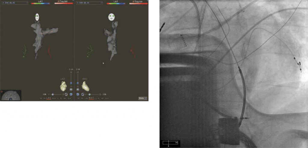
A 53 year-old man was chronically followed by our team for ischemic cardiomyopathy, ejection fraction of 10% and a right-sided biventricular Biotronik defibrillator. Additional comorbidities included high blood pressure, diabetes and renal failure with an indwelling dialysis port in the left-sided subclavian vein. He was emergently admitted to ICU after having episodes of syncope confirmed to be secondary to ventricular fibrillation. His device interrogation revealed three ineffective shocks at maximum output, with effective late conversion on the final 4th shock although spontaneous termination of arrhythmia could not be excluded. Once he recovered from the critical event a defibrillator threshold testing was performed in the EP lab. Unfortunately, despite trying several configurations and maximum defibrillation output all internal shocks failed requiring external defibrillation. With his device placement, there was a concern for ineffective defibrillator vectors between the ICD can in the right subclavian area and the distal RV coil at the opposite diameter in the left thorax at the cardiac apex. In the setting of high defibrillator threshold, the upgrade options include placing a subcutaneous array, a coronary sinus coil or an azygos coil. Since our patient had a right sided biventricular ICD, the only option available was placing an azygos coil for an optimized defibrillator threshold with direct anterior- posterior vector. While placing an azygos coil from the left side represents a procedure of average difficulty, it is most frequently performed from a left sided access allowing a natural trajectory of the delivery sheath and lead into the azygos vein. In order to avoid a potentially diffisituation with the right sided access in this case, we decided to perform a CT angiogram of the superior vena cava and tributaries including the azygos veins was obtained pre-procedure (Figure 1). We then brought the patient the EP laboratory, opened the pocked and obtained access in the right axillary vein with a standard approach. A slight stenosis was dilated with incremental dilators and sheaths until we were able to place a short 8 Fr peel away sheath. Following sheath insertion, we advanced a decapolar mapping catheter (Decanav Biosense Webster®) and created a 3D geometry of the superior vena cava and subclavian vein (Figure 2). In order to locate the azygos vein take off with the mapping catheter, we merged the 3D map with the previously acquired CT angiogram (Figure 3). Once the overlapping images were synchronized, we easily advanced the decanav catheter in the azygos vein with a subsequent advancement of the 8 Fr sheath over it. The last stage of the procedure consistent of inserting the additional coil into the azygos vein through the existing sheath (Figure 4). Defibrillator thresholds were performed and were found to be effective at 20 Joules below the maximum device output.

Figure 1. CT angiogram representing the superior vena cava, right atrium, tricuspid valve and inferior vena cava. Note the brown catheter placed in the azygos vein clearly identified on the CT angiogram.

Figure 2. Three dimensional map of the right subclavian vein, superior vena cava and azygos vein obtained during the case with the decapolar mapping catheter.
Figure 4. Chest X ray showing the placement of the azygos lead behind the left ventricle and to the left of the spine. The left ventricle is “sandwiched between the right ventricle apical lead and the azygos coil advanced below the diaphragm. The LV quadripolar lead is visualized in a postero-lateral location.

Figure 3. Overlap of the 3D images acquired with CT angiogram and 3D mapping with clear delineation of the azygos origin and a road map for placement of the mapping catheter.
DISCUSSION
We present a challenging case of successful RV coil insertion in the azygos vein via difficult right-sided access utilizing 3D mapping and merging with CT angiography. While 3d mapping overlapping a previously acquired CT is a technique already employed in atrial fibrillation or ventricular tachycardia ablations, we report an off label use of a 3 d mapping system for azygos vein cannulation and ICD upgrade. This procedure would have been difficult if not impossible with fluoroscopy alone and importantly, the patient’s prognosis without this procedure would have been less than 6 months survival. At follow-up, impedance in the azygos coil was measured at 65-68 ohm while the patient continues to be protected from sudden cardiac death due to an effective defibrillating system in place. To our knowledge, the use of 3D mapping has not been previously used in a similar set up and may open the door for future procedures sans fl uoroscopy like His bundle pacing and LV lead placement in complex CS anatomy.
Conflict of interest: none declared.
References
1. Rahaby, M, Niazi, I. Azygous vein coil: bailout strategy for high defi-brillation thresholds. Journal of innovations in cardiac rhythm man-agement 2012.
2. Moran, DP, Bhutta, U, Yearoo, I, Keelan, E, O’Neill, J, Galvin, J. Case report of an anomalous single azygous venous coil insertion to re-duce the defibrillation threshold in a patient with a right-sided delto-pectoral ICD implant. Heart Asia 2013, 5(1): 28/29.
 This work is licensed under a