Radu Miftode1,2, Adrian Pintilie1, Andreea Timofte1, Ana-Maria Statescu1, Cipriana Stefanescu1,2, Mihai Gutu1, Ionut Tudorancea1,2, Irina Iuliana Costache1,2*, Antoniu Octavian Petris1,2
1 „Sf. Spiridon” County Emergency Hospital, Iasi, Romania
2 „Grigore T. Popa” University of Medicine and Pharmacy, Iasi, Romania
Abstract: Background – The use of stress myocardial perfusion scintigraphy, Single-photon Emission Computed To-mography (SPECT), is nowadays revived in the evaluation of patients with suspected ischemic heart disease in order to indi-rectly assess blood flow and myocardial fl ow reserve. Aim of the study – To identify clinical, electrocardiographic (ECG) and echocardiographic features associated with SPECT abnormalities in myocardial perfusion. Materials and methods – We conducted an observational, prospective study, on 17 successively enrolled patients, 58.83% men, aged 35-79 years (57.47+/-13.18 years), admitted in a cardiology clinic of an academic, general, non-coronarography capable hospital, where every resource to identify and quantify myocardial ischemia must be used. The following data was collecting: cardiovascular risk factors, myocardial ischemia on ECG, echocardiographic quantification of cardiac chamber size and left ventricular ejection fraction (LVEF) and perfusion defects on stress myocardial SPECT. Results – Perfusion defects on 99mTc-MIBI SPECT were detected on the majority of patients (76.47%) while ECG was suggestive for ischemia in only 52.94% of the total included patients. There were no signifi cant differences on the cardiovascular risk factors between the subgroup of patients with or without defects in myocardial perfusion SPECT. Anterior wall perfusion defects have been closely and di-rectly correlated with right ventricular telediastolic diameter (p < 0.05, r = 0.730) and indirectly with BNP level (p < 0.01, r = -0.891) and inferior vena cava diameter (p < 0.05, r = -0.651). Lateral wall perfusion defects have been closely and di-rectly correlated with creatinkinase, CK-MB level (p < 0.01, r = 0.711; p < 0.05, r = 0.607, respectively) and left ventricular posterior wall thickness (p < 0.01, r = 0.765) and indirectly with LVEF-echo (p < 0.051, r = -0.498). Inferior wall perfusion defects have been closely and directly correlated with BNP level (p < 0.05, r = 0.735) and indirectly with smoker status (p < 0.01, r = -0.683). Conclusion – We provide an early insight into SPECT parameters versus ECG/ echocardiographic in patients with IHD assessed in Northeastern Romania, revealing some clinical, ECG and echocardiographic features, opening the perspectives for a larger prospective study.
Keywords: cardiovascular disease, SPECT, myocardial scintigraphy, ischemia, perfusion.
INTRODUCTION
Cardiovascular disease (CVD) is the leading cause of death worldwide, accounting for 31.5% of all deaths, with ischemic heart disease (IHD) and stroke being the most frequent1. The incidence of IHD is increasing, with 1.6% of the adult population (112 million peo-ple) suffering from ischemic heart disease, men having a slightly higher prevalence (1.7%)2. In the European region, CVD causes more than 4 million deaths each year, accounting for 45% of all deaths. IHD and cere-bro-vascular disease were the most common causes of CVD deaths, with a total number of 1.8 million and 1.0 million deaths, respectively1. Romania has one of the highest mortality rates in Europe, CVD being the main cause of death (62.1%), with a third of deaths caused by IHD alone3. Thereby, IHD is not only a me-dical aspect, but its approach also involves economic challenges (high burden on the public health system), social issues (poor quality of life for the patients and their relatives) and even some ethical problems4. Myo-cardial perfusion scintigraphy is a non-invasive cardiac imaging test that uses electromagnetic gamma radi-ation to obtain images in order to indirectly assess blood flow and myocardial flow reserve, with a key role in the diagnosis and severity assessment of IHD in patients with suspected or known coronary artery pathology5,6. The objective of the present study was to identify the risk factors associated with abnormalities in myocardial perfusion and to establish correlations between the ischemic electrocardiography changes and affected coronary territory.
METHODS
We conducted an observational study, which included 17 patients, 18 men and 9 women, age range 35-79 years (57,47 +/- 13,18 years), admitted at the Car-diology Clinic of „Sf. Spiridon” Clinical Emergency County Hospital Iasi (hospital without coronary angio-graphy facilities), between January 2016 and June 2018. The following data was collected: history of hyperten-sion, dyslipidemia, smoking, obesity, diabetes, chronic kidney disease, family history or cardiovascular disea-se, electrocardiographic modifi cations suggestive for myocardial ischemia, quantification of cardiac chamber size and left ventricular ejection fraction (LVEF) on echocardiography (GE Vivid 7 Ultrasound Machine®), and perfusion defects on stress myocardial scintigra-phy (CardioSPECT DIACAM – Siemens®).
In terms of inclusion criteria, patients enrolled in the study:
- presented at admission at least one of the following signs or symptoms: chest pain, dyspnea or other anginal equivalents;
- showed initial or subsequent electrocardiogra-phic modifications suggestive of myocardial is-chemia;
- were previously diagnosed with IHD or acute co-ronary syndromes (documented in their medical records).
The exclusion criteria were:
- patients with severe cardiac pathologies (acute myocardial infarction, unstable angina, sympto-matic cardiac arrhythmias with hemodynamic changes, severe aortic stenosis, recent pulmonary thromboembolism, acute myocarditis / peri-carditis and aortic dissection);
- patients who refused a complete preliminary eva-luation (clinical exam, ECG, Echocardiography, blood samples);
- patients with particular conditions that preven-ted a correct scintigraphic examination, especi-ally at effort (surgery, extreme obesity, severe osteo-articular pathology etc);
- patients with neuro-psychiatric pathologies or special socio-cultural beliefs who disagreed with the standard written consent.
Prior to the study, drugs that could have infl uenced the results were suppressed: beta blockers (72 hours), calcium channel blockers (48-72 hours), long-acting ni-trates (12 hours).
The myocardial perfusion scintigraphy was perfor-med using the Siemens E. Cam Signature Series Dual Detector® with variable angle + cardio. For the scin-tigraphic examination, the 99mTc-MIBI radiopharma-ceutical was used in a one day or two days protocol, both at rest and with stress test (Bruce and modified Bruce protocol). The one-day protocol consisted of the injection of 8 mCi for stress test in peak effort (maximum theoretical heart rate/ 220 – age) followed by a 3 hours pause and a subsequent 24 mCi adminis-tration at rest. In the case of the two-days protocol, in the first 24 hours the physical stress test was carried out with the acquisition of gated, synchronous ECG images 30 minutes after the injection, while in the se-cond day, the protocol was performed at rest, with the acquisition of gated, synchronous ECG at 60 mi-nutes after injection7,8. Stress test termination criteria were: severe ST segment depression (>3 mm), ST seg-ment elevation >1 mm in leads without pre-existing Q waves due to prior myocardial infarction, sustained ventricular tachycardia (VT) or other arrhythmias, in-cluding second or third-degree atrioventricular block, severe chest pain, dyspnea or confusion, decreased systolic blood pressure (a drop >20 mm), increased blood pressure (systolic >300 mm, diastolic >130 mm), signs of poor peripheral perfusion (cyanosis or pallor), the patient’s request to stop the test9.
The image processing was performed with QGS software, using three sections: short axis (SA), vertical long axis (VLA) and horizontal long axis (HLA). For the three-dimensional representation of the left ven-tricle and the evaluation of left ventricular function, the ECG-gated SPECT technique (image processing synchronized with the electrocardiogram) was used. Myocardial perfusion scintigram was analyzed by two senior doctors in nuclear medicine using the semi-quantitative visual assessment (17 segments model) and also a quantitative assessment (bull’s eye). A five point scoring per myocardial segment allows the calcu-lation of a series of scores: summed stress score (SSS), summed difference score (SDS) and summed rest sco-re (SRS), which can be used to represent the global indices of myocardial perfusion7. Myocardial perfusion was considered normal when the tracer’s distributi-on was homogeneous both at stress and at rest. The presence of a reversible perfusion defect (low uptake, reversible at rest) was suggestive for myocardial ische-mia. Myocardial infarction with adjacent ischemia was associated with the occurrence of a partially reversi-ble perfusion defect (low uptake, partially reversible at rest), while a fixed perfusion defect (low uptake persistent at rest) occurred in the presence of an area of necrosis (myocardial infarction) or sclerosis (sequel of myocardial infarction)8.
Ethical aspects
The participants were informed about the subject, purpose and rules of the study. Each participant signed and agreed on admission to participate in the research process, their data being processed anonymously.
Statistical analysis
All data was statistically analyzed using the SPSS v24 for Windows (SPSS Inc., Chicago, IL, USA), Microsoft Ex-cel 2003 (1985-2003 Microsoft Corporation®) softwa-re, univariate statistical analysis (frequency, mean, ran-ge, and median) and comparison test being performed variables Chi2, Student’s t test comparing two means (quantitative). Data was expressed as mean ± SD, while for p-value we were using the two-tailed test.
RESULTS
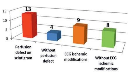
Out of a total of 17 patients, 10 (58.83%) were men. Perfusion defects on scintigram were detected on the majority of patients (76.47%) while, for comparison, the ECG was suggestive for ischemia in only 52.94% of the total included patients (Figure 1).
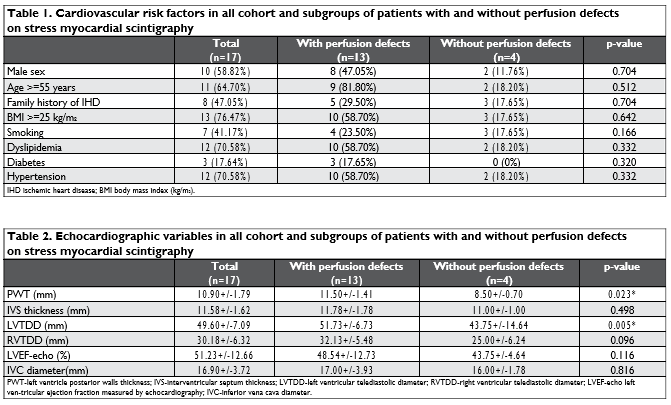
We assessed the incidence of some of the most important cardiovascular risk factors in the two subgroups (patients with or without perfusion defects on scintigram). There were no significant differen-ces on some cardiovascular risk factors (age above 55, male sex and body mass index (BMI) >25 kg/m2) between the subgroup of patients with or without de-fects in myocardial perfusion scintigraphy (Table 1). Smoking status (41.15% of total patients), presented a roughly similar proportion of perfusion defects and normal scintigrams (23.5% vs 17.65%, p=0.127).
We found no significant difference between ejec-tion fraction assessed by echocardiography (51.23+/-12.66%, median 55%) or by myocardial scintigraphy (53.24+/-14.74%, median 55%) (p=0.118). Among the echocardiographic variables which were assessed, PWT and left ventricular ejection fraction showed significant differences between the two subgroups analyzed, with the p value of 0.023 and 0.005, respec-tively (Table 2). The left ventricular ejection fraction has been closely (p<0.005) and directly correlated with the presence of dyslipidemia (r = 0.664), maintai-ning sinus rhythm (r = 0.735) and fibrinogen levels (r = 0.620) (r = Pearson correlation, sig. 2-tailed).
We have noticed that a reduced left ventricle ejec-tion fraction on scintigraphy (LVEF-scin) is correlated with the presence of perfusion defects (p=0.05), while when this parameter was assessed using echocardiography such significant correlation can not be ascer-tained (p=0.218). From the entire lot, only 3 patients (17.65%) have a dilated LV >55 mm, this data infirming any relationship between the LV dimension and an op-timal scintigraphy perfusion in this study (LV range 39-67 mm, mean 49.3+/-7.1 mm, p=0.304). The presence of aortic atherosclerosis, a marker of severity, is highly associated with perfusion abnormalities, because 9 out of 11 patients with aortic atherosclerosis had also per-fusion defects on scintigraphy (p=0.047) (Table 3).
With regard to ECG modifications, although we have noticed certain differences between the two subgroups, they have failed to reach the statistical significance threshold, both for ischemia (p=0.661) and arrhythmias (p=0.494). In terms of cardiac bio-markers, although we have noticed the association of perfusion defects in patients with elevated levels of Troponin I or NT-pro BNP, these values failed to re-ach the significance threshold, with a p-value of 0.326 and 0.895, respectively.
Anterior wall perfusion defects have been closely and directly correlated with right ventricular end-dias-tolic diameter (p <0.05, r = 0.730) and indirectly corre-lated with BNP level (p <0.01, r = -0.891) and inferior vena cava diameter (p <0.05, r = -0.651). Lateral wall perfusion defects have been closely and directly cor-related not only with creatinkinase and CK-MB levels (p <0.01, r = 0.711; p <0.05, r = 0.607, respectively) but also with left ventricular posterior wall thickness (p <0.01, r = 0.765), while an indirect association was observed with echo-determined LVEF (p <0.051, r = -0.498). Inferior wall perfusion defects have been clo-sely and directly correlated with BNP level (p <0.05, r = 0.735) and indirectly with the smoking status (p <0.01, r = -0.683).
There are no significant gender differences regar-ding perfusion defects on scintigram (p=0.683) or is-chemic alterations on ECG (p=0.778). Both men and women presented hypertension in similar proportions (7 men out of 10 vs 5 women out of 7, p=0.951), while concerning the lipidic profile, we noticed a small diffe-rence between genders, but without statistical signifi-cance (p=0.323). Only in terms of hemoglobin there is a difference between the two genders (p=0.013).
We have identified some considerable differences between patients with age above or under 55 years regarding the family history of IHD (p=0.008), hyper-tension (p=0.010), serum levels of CK (p=0.033), LDL-C (p=0.030), HDL-C (p=0.036) and creatinine clearance (p=0.004).
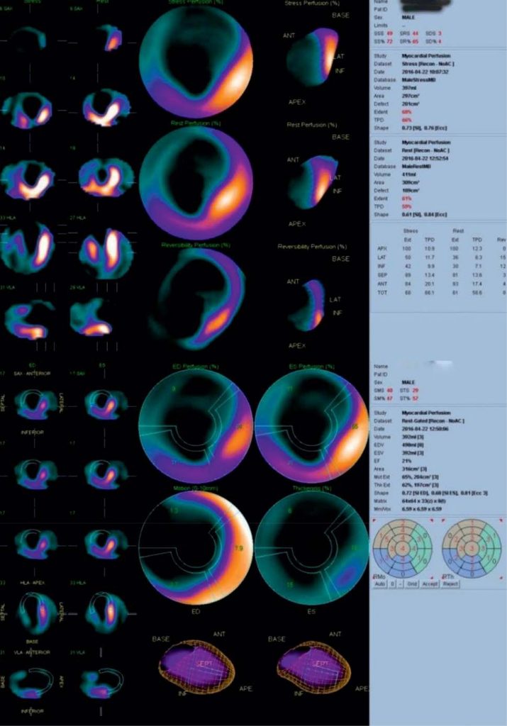
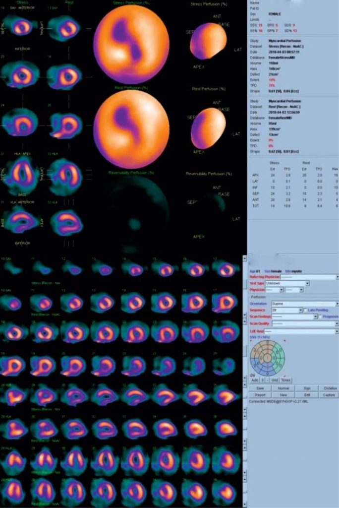
Figures 2 and 3 show two examples of changes re-garding the defects of myocardial perfusion in specific clinical situations, as highlighted by scintigraphic examination.

Figure 1. Frequency of patients with ischemic modifications – ischemia on ECG and perfusion defects on stress myocardial scintigraphy.


Figure 2 A) Myocardial perfusion scintigraphy detect a large fixed perfusion defect, involving left ventricular apex, half of the septum, and almost all the anterior wall. The defect corresponds to post-myocardial infarction scars with left ventricular aneurysm. At rest, a medium defect is recovered from the inferolateral portion. B) In the gated sequence, synchronized with the ECG, lower thickness of the myocardial wall is observed in the same extended area (images from the Archive of the Nuclear Medicine Laboratory, Sf. Spiridon Hospital, Iaşi).

Figure 3. Myocardial perfusion scintigraphic assessment: SSS = 11, SS = 16%, SD = 13 scores indicate a high probability of effort induced ischemia. The severity of the previous perfusion defect suggests significant stenosis of left anterior descending artery (LAD) (images from the Archive of the Nuclear Medicine Laboratory, Sf. Spiridon Hospital, Iasi).
DISCUSSIONS
Albeit severe ischemia in a coronary territory causes a number of functional and clinical changes (ischemic cascade), 50% of patients with IHD may have normal findings on resting ECG, if the left ventricular function is preserved10. Recommendations for diagnostic tes-ting need to take into account the pre-test probability, the major determinants being: age, gender and the na-ture of symptoms. Other variations can occur due to the interdependence between clinical likelihood that a given patient will have IHD and the performance of the available diagnostic methods (ranging from a sen-sitivity of 45-50% for exercise ECG to 95-99% for co-ronary computed tomography angiography and a spe-cificity of 85-90% for exercise ECG and 64-83 % for coronary computed tomography angiography)11. The use of myocardial perfusion scintigraphy is revived nowadays in the evaluation of patients with suspec-ted ICD due to its high diagnostic accuracy, as well as being able to defi ne the extent, severity and location of myocardial perfusion abnormalities.12 In myocardial perfusion scintigraphy, certain radiotracers are injec-ted during the procedure and, due to affi nity for the myocardium, they bring information concerning both perfusion and metabolic functional status13. A normal resting myocardial perfusion scan virtually excludes major myocardial infarction, while an abnormal acute scan indicates the presence of IHD and the need for further investigations.
We conducted an observational, prospective study, which included 17 patients, 58.83% men, aged 35-79 years (57.47+/-13.18 years), admitted in a cardiology clinic from an academic, general, non-coronarography capable hospital, where every resource to identify and quantify myocardial ischemia must be used. Remem-ber that in Romania the reported number of inter-ventional cardiologists per million people is only 4.414.
We did not found any significant differences in terms of cardiovascular risk factors between the subgroup of patients with (13 cases) or without (4 cases) proven defects in myocardial perfusion scintigraphy. Howe-ver, compared to our data collected from a small lot, a large study conducted by De Lorenzo et al, comprising more than 4000 patients, highlighted the strong asso-ciation between diabetes and perfusion defects, but no important differences in terms of long-term mortality, compared to non-diabetic patients with IHD15. Some different results were found by Berman et al, who ob-served that diabetic patients with perfusion defects had an increased risk of cardio-vascular events, espe-cially if insulin treatment was required16. Regarding the gender infl uence, the same study revealed that women presented lower and smaller perfusion defects than men, but women with diabetes were at greater risk of adverse outcome for any level of stress perfusion defects compared with non-diabetic women or even with diabetic men16. We didn’t noticed any signifi cant difference between genders regarding perfusion de-fects on scintigram (p=0.683) or ischemic alterations on ECG (p=0.778). However, we highlighted 2 cases with ST-T modifications on ECG but without perfusi-on defects at scintigraphy, both patients being females. Of course, this situation can occur due to technically false-positive ECG or false negative scintigraphy, but an explanation given by some authors is the vasocon-strictor, digoxin-like effect of estrogen during physical exercise, that can cause ECG changes, without or only with minimal diffuse modifications on scintigram17,18,19. Another hypothesis for the discordance between ECG findings and scintigram is the presence of a small heart, typically found in women, which can cause difficulties for a gamma-camera with limited spatial resolution20. Thereby, it is worth mentioning that in our study, the LV dimensions were below 45 mm in both cases with ECG vs. scintigraphy discordance, sustaining the abo-ve-mentioned theory. From the other perspective, an increased LV diameter >55 mm has been associated in many studies not only with important ECG changes, but also with perfusion defects, suggesting a poor out-come in these patients21.
Evaluation of the ejection fraction by echocardio-graphy and scintigraphy did not reveal any significant differences for all cohort (51.23+/-12.66% vs 53.24+/-14.74%, p=0.118), but among the echocardiographic variables assessed, PWT and LVEF showed significant differences between the two analyzed subgroups, with a p value of 0.023 and 0.005, respectively. We obser-ved that all patients with a reduced LVEF determi-ned by scintigraphy also presented perfusion defects (p=0.05), further confirming the bidirectional relation-ship between an altered myocardial blood flow and a reduced contractility. However, there is controversy about the best method to determine LVEF in patients with CAD, with scintigraphy method sometimes ove-restimating the contractility compared to echocardi-ography, especially in stress condition, as shown in a study conducted by Godkar et al.22 No ischemic mitral regurgitation was observed in the analyzed group, ne-ither at rest nor in the stress23. Although there is scarce data in literature referring to the cardiac enzymes or biomarkers levels in pati-ents undergoing perfusion scintigraphy, some authors considered that a high serum-level of NT pro-BNP hi-ghly correlates with perfusion defects with a specificity >95%, even proposing its use as a screening method and a predictor of a normal scintigram24. In our study we observed that almost all patients with an elevated troponin I (>0.020 ng/mL) also presented perfusion defects. Anterior wall perfusion defects have been closely and indirectly associated with BNP level (p <0.01, r = -0.891), lateral wall perfusion defects have been closely and directly correlated with creatinkinase and CK-MB level (p <0.01, r = 0.711; p <0.05, r = 0.607, respectively) and inferior wall perfusion defects have been closely and directly correlated with BNP level (p < 0.05, r = 0.735).
CONCLUSION
This article provides an early insight into use of the perfusion scintigraphy in patients with IHD from Northeastern Romania, revealing some clinical, elec-trocardiographic and echocardiographic features, ope-ning the perspectives for a larger prospective study.
Study limitations
The limitations of this study included a small lot of analyzed patients, lack of details regarding the durati-on of ischemia prior to hospitalization and the absence of some patient’s full medical data records to attest other unspecified cardio-vascular comorbidities or other previous treatments or explorations. Another limitation of the study was determined by the absence of an immediately available coronary angiography in the Clinic, for a more accurate, synergic assessment of the ischemic territory. In addition, some of the hospitalized patients with IHD and potential positive perfusion defects at scintigraphy, have refused the ex-ploration.
Acknowledgments: We thank the team of the Nuclear Medicine Laboratory I – Radioisotopes led by Prof. Dr. Cipriana Ştefănescu. Thanks to Dr. Adrian Pintilie and Dr. Andreea Timofte who collected the data.
Conflict of interest: None declared.
References
1. Townsend N, Wilson L, Bhatnagar P, Wickramasinghe K, Rayner M, Nichols M. Cardiovascular disease in Europe: epidemiological update 2016. Eur Heart J. 2016; 37(42): 3232-3245.
2. Vos T, Flaxman AD, Naghavi M, et al. Years lived with disability (YLDs) for 1160 sequelae of 289 disease and injuries 1990-2010: a systematic analysis for the Global Burden of Disease Study 2010. Lancet 2012; 380(9859): 2163-2196.
3. Vlădescu C, Scîntee G, Olsavszky V, Allin S, Mladovsy P. Romania: Health system review. Health systems in Transition, 2008; 10(3): 1-172.
4. Petriş A, Cimpoesu D, Costache I, Rotariu I. Do not resucitate de-cision (I). Ethical issues during cardiopulmonary resuscitation. Rev Rom de Bioetică 2011; 9(2): 40 – 49.
5. Russe RR, Zaret BL. Nuclear cardiology: Present and future. Curr Probl Cardiol 2006; 31: 557-629.
6. Hendel RC, Berman DS, Di Carli MF, et al. ACCF/ASNC/ACR/ AHA/ASE/SCCT/SCMR/SNM 2009 appropriate use criteria for car-diac radionuclide imaging: A report of the American College of Car-diology Foundation Appropriate Use Criteria Task Force, the American Society of Nuclear Cardiology, the American College Radiology, the American Heart Association, the American Society of Echocar-diography, the Society of Cardiovascular Computed Tomography, the Society for Cardiovascular Magnetic Resonance, and the Society of Nuclear Medicine, endorsed by the American College of Emer-gency Physicians. Circulation 2009; 119: e561-87.
7. Fathala A. Myocardial perfusion scintigraphy: techniques, interpretation, indications and reporting. Ann Saudi Med 2011; 31(6): 625-34.
8. Ştefănescu C, Rusu V. Scintigrafia de perfuzie miocardică, 306-313. De la fizica şi biofizica radiofarmaceuticelor la imagini funcţionale şi moleculare, Ed. Tehnopress, Iaşi, 2007. ISBN 973-702-495-8.
9. Fletcher GF, Ades PA, Kligfield P, et al. American Heart Association Exercise, Cardiac Rehabilitation, and Prevention Committee of the Council on Clinical Cardiology, Council on Nutrition, Physical Activ-ity and Metabolism, Council on Cardiovascular and Stroke Nursing, and Council on Epidemiology and Prevention. Exercise standards for testing and training: a scientific statement from the American Heart Association. Circulation. 2013; 128(8): 873-934.
10. Harris PJ, Behar VS, Conley MJ, et al. The prognostic significance of 50% coronary stenosis in medically treated patients with coronary artery disease. Circulation 1980; 62: 240-248.
11. Montalescot G, Sechtem U, Achenbach S, et al. 2013 ESC guidelines on the management of stable coronary artery disease. Eur Heart J 2013; 34: 2949–3003.
12. Smanio PEP, Silva JH, Holtz JV, Ueda L, Abreu M, Marques C, Mach-ado C. Myocardial scintigraphy in the evaluation of cardiac events in patients without typical symptoms. Arq Bras Cardiol 2015; 105(2): 112–122.
13. Hendel RC, Abbott BG, Bateman TM, Blankstein R, Calnon DA, Lep-po JA, et al; American Society of Nuclear Cardiology. The role of ra-dionuclide myocardial perfusion imaging for asymptomatic individu-als. J Nucl Cardiol 2011; 18(1): 3-15.
14. Timmis A, Townsend N, Gale C, Grobbee R, Maniadakis N, Flather M, Wilkins E, Wright L, Vos R, Bax J, Blum M, Pinto F, Vardas P. European Society of Cardiology: Cardiovascular Disease Statistics 2017. Eur Heart J 2018; 39: 508–577.
15. De Lorenzo A, Souza VF, Glerian L, Lima RS. Prognostic Assessment of Diabetics Using Myocardial Perfusion Imaging: Diabetes Mellitus is Still a Coronary Artery Disease Equivalent. Open Cardiovasc Med J.
2017; 11: 76-83.
16. Berman DS, Kang X, Hayes SW, Friedman JD, Cohen I, Abidov A, Hachamovitch R. Adenosine myocardial perfusion single-photon emission computed tomography in women compared with men: Im-pact of diabetes mellitus on incremental prognostic value and effect on patient management. Journal of the American College of Cardiol-ogy 2003; 41(7): 1125-1133.
17. Tremmel JA, Yeung AC. Ischemic heart disease in women: an appropriate time to discriminate. Rev Cardiovasc Med 2007; 8: 61–68.
18. Azemi T, Rai M, Parwani P, Baghdasarian S, Kazi F, Ahlberg AW, et al. Electrocardiographic changes during vasodilator SPECT myocar-dial perfusion imaging: does it affect diagnosis or prognosis? J Nucl Cardiol 2011; 19:84–91.
19. Bokhari S, Bergmann SR. The effect of estrogen compared to estro-gen plus progesterone on the exercise electrocardiogram. J Am Coll Cardiol 2002; 40: 1092–1096.
20. Taywade SK, Ramaiah VL, Basavaraja H, Venkatasubramaniam PR, Selvakumar J. Prevalence of ECG changes during adenosine stress and its association with perfusion defect on myocardial perfusion scintigraphy. Nuclear Medicine Communications 2017; 38(4): 291-29.
21. Singh P, Bhatt B, Pawar SU, Kamra A, Shetye S, Ghorpade M. Role of myocardial perfusion study in differentiating ischemic versus non-ischemic cardiomyopathy using quantitative parameters. Indian Jour-nal of Nuclear Medicine 2018; 33(1): 32.
22. Godkar D, Bachu K, Dave B, Megna R, Niranjan S, Khanna A. Com-parison and co-relation of invasive and noninvasive methods of ejec-tion fraction measurement. J Natl Med Assoc. 2007; 99(11): 1227-8: 1231-4.
23. Petriş AO, Iliescu D, Alexandrescu DM, Costache II. Ischemic mitral regurgitation in patients with acute myocardial infarction. Rev Med Chi Soc Med Nat Iaşi 2014; 118: 618-623.
24. Rathcke CN, Kjøller E, Fogh-Andersen N, Zerahn B, Vestergaard H. NT-proBNP and circulating inflammation markers in prediction of a normal myocardial scintigraphy in patients with symptoms of coronary artery disease. PLoS One 2010; 5(12): e14196.
 This work is licensed under a