Ioan Tiberiu Nanea1
1 Department of Cardiology and Internal Medicine, „Prof. Dr. Theodor Burghele” Clinical Hospital, „Carol Davila” University of Medicine and Pharmacy, Bucharest, Romania
INTRODUCTION
Notwithstanding the multimodality myocardial ima-ging age, the information obtained by analyzing the electrocardiographic changes that occur during the acute coronary occlusion is underestimated.
The dynamics of ST segment elevation is essenti-al for diagnosis, therapeutic approach and efficiency, and also for risk stratification of the patient with acute myocardial infarction. Many studies have tried to esti-mate the myocardial area at risk and eventually the es-tablished myocardial infarction, by ascertaining a score of the ST segment magnitude and of the number of electrocardiographic leads where it happens.
Thus, in case of anterior myocardial infarction the number of leads with ST segment elevation has been strong correlated with the infarcted myocardium, and in case of inferior one the magnitude of ST segment elevation has been related to the final size of the in-farction.
The size and extension of the ST segment elevation is also correlated with the presence of an effective collateral circulation and with a residual flow through the involved coronary artery which is adequate to the viability of the subjacent tissue.
In patiets who benefited from efficient coronary an-gioplasty there is a poorer correlation between these electrocardiographic scores and the myocardium at risk identified by studies with radionuclides or by mag-netic resonance imaging. However, the assessment of the myocardium at risk using the cuantifi cation of the magnitude and extension of ST segment elevation is an independent method which is different from other technics performed with the same purpose. Therefore, the analysis of the ST elevation particularities stands up-to-date in establishing the myocardial at risk and in evaluation of the prognosis to the patient with acute coronary occlusion.
THEORIES EXPLAINING THE ST SEGMENT ELEVATION
Information regarding therapeutic efficacy, prognosis and risk stratification in the patients with acute myo-cardial infarction, obtained by analysing the electro-cardiographic changes is underestimated.
The dynamic ST segment elevation represents the essential change in the matter of diagnosis (STEMI), complications, therapeutic approach, therapeutic effi – cacy and risk stratification in patients with acute coro-nary occlusion1.
The myocardial lesion, expressed through ST seg-ment elevation or depression, denotes an advanced stage of myocardial ischemia, signifying transmural le-sion with a subepicardial prevailing pattern.
There are two theories that explain the ST segment elevation: diastolic and systolic current of injury.
During the electric diastole (TQ interval), the inju-red myocardium remains depolarized, in contrast to the normal adjacent myocardium which becomes re-polarized. Thus, a potential difference appears betwe-en these zones, a diastolic current of injury whose vector is oriented from the injured zone towards the normal electropositive myocardium, determining a TQ segment depression. The superfi cial electrocar-diogram records the TQ segment depression as ST segment elevation, as electrocardiographs use an am-plified current which adjustes in a proportional mode every TQ segment depression.
During the electric systole (QT interval) a potential difference (systolic current of injury) appears between the injured zone which is early repolarized and the normal adjacent depolarized zone. The vector of the systolic current of injury is oriented towards the elec-tropositive injured zone. The superfi cial electrocar-diogram records a ST segment elevation. The leads which are oriented to the injured zones will record a ST segment elevation, and the opposed ones a ST segment depression.
The magnitude of the ST elevation is measured at J point (the junction between the end of the QRS and the beginning of the ST segment), having as orientation mark the TP segment which is isoelectric.
The morphological aspects, the dinamics, the size and the number of electrocardiographic leads where there is recorded the ST segment elevation offer in-formation regarding:
-
-
- Diagnosis and myocardial area at risk of develo-ping subsequent myocardial necrosis (analysing the ST segment magnitude and the number of the involved leads).
- Risk stratifi cation in patients with acute myocar-dial infarction (analysing the morphology and the leads with ST segment elevation).
- Efficacy of reperfusion therapy (analysing the ST segment dinamics).
- Identification of the reperfusion injury (noticing the ST segment dinamics).
- Localization of the myocardial necrosis (analysing the ST segment location in the electrocardiogra-phic leads).
- Identification of the coronary artery responsible of occlusion (measuring the magnitude and loca-lizing the ST segment elevation on electrocardi-ogram).
- Evaluation of the residual flow (measuring the size of ST segment elevation).
- Identification of ventricular aneurysm (analysing the electrocardiographic signs of necrosis and lesion).
- Prediction of left ventricular function (existence of ST segment elevation in more leads is associ-ated with reduced ejection fraction).
- Diagnosis and myocardial area at risk of develo-ping subsequent myocardial necrosis
The dinamics of the ST segment elevation after co-ronary occlusion is influenced by:
a.Duration of the occlusion
b.Antegrade residual flow through the coronary artery responsible for infarction;
c.Myocardium at risk of developing an established myocardial infarction;
d.Flow level through the coronary vessels which are collateral to the injured area;
e.Ischemic preconditioning;
f.Myocardial oxygen consumption;
g.Physiological fibrinolysis;
h.Moment of performing the reperfusion therapy;
i.Quality of reperfusion.
Many studies suggested1 that the level of initial mag-nitude of the ST segment elevation (at the beginning of the coronary syndrome) and its extension in more leads can anticipate the area of myocardial necrosis (the final risk of necrosis). It is considered that if the injured myocardial area increases, the transmural elec-trical potential differences between the ischemic area and the normal myocardium will be augmented, cau-sing ST segment elevation2,3. According to this theory, the magnitude of the ST segment elevation reflects the extension of myocardium at risk that will necrosed afterwards. The initial ST segment elevation at the ad-mission has predective value for the estimation of the myocardial infarction size later established.
Many studies tried to estimate the myocardial area at risk, the size of myocardial infarction, developing a score using the value of the ST segment magnitude and the number of electrocardiographic leads where it occured. Aldrich has established formulas for evalu-ation of the final myocardial infarction size taking into account the anterior and inferior localization4.
Formula for anterior myocardial infarction = 3(1.5 [number of leads with ST elevation – 0.4])
Formula for inferior myocardial infarction = 3(0.6 [ ST elevation in leads II, III, aVF] + 2.0).
The final size of myocardial infarction was estima-ted through QRS score described by Selvester, which was calculated at discharge5. Selvester score for eva-luation of the myocardial infarction size consists of 57 criteria based on the duration of Q or R waves, the amplitude of the R or S waves and the amplitude of the R/Q or R/S ratio. Each point in the Selvester score corresponds to 3% of the myocardial infarction size. The Selvester QRS Score for evaluation of the myocardial infarction size was validated by morphopathological studies (by measuring of the myocardial infarction quantity during autopsy) and by radionuclide studies5,6 (Figure 1).
In a study performed by Aldrich on 148 patients (without reperfusion treatment), the number of leads with ST segment elevation that signifies the myocardi-al infarction at risk was correlated with the size of the established anterior myocardial infarction (r = 0.72). In contrast, in case of inferior infarction, the sum of ST elevations magnitude was strongly correlated to the size of the established myocardial infarction (r = 0.61)4.
In his study which included 67 patients with coro-nary angioplasty, Thymothy estimated the size of the myocadial infarction at risk electrocardiographically assessed, by comparison with radionuclide and angio-graphic measurements. For the calculus of the area at risk, it was used a different formula compared to the one described by Aldrich. So, for the anterior myocar-dial infarction, the formula becomes:
4.5 x (number of leads with ST elevation ≥ 1 mm) – 1.2;
and for the inferior myocardial infarction:
1.8 x ( ST elevations in mm in leads II, III, aVF) + 6.6.The myocardial area at risk and the collateral cir-culation were evaluated by radionuclide studies quan-tifying the perfusion defects, and by coronary angio-graphy. The collateral coronary circulation was evalu-ated, using Rentrop score.7 The authors conclude that the ST segment elevation score is correlated to the radionuclide or to the coronary angiographic evaluati-on of the flow through the collateral coronary circula-tion. The magnitude of the ST elevation (myocardium at risk) has a poor correlation to the established myo-cardial infarction detected by perfusion defects.4 How-ever, in many cases we obtained a good correlation between the magnitude of the ST segment eleveation and the size of established myocardial infarction de-tected by perfusion defects (Figure 2).
In patients who received coronary reperfusion therapy, there is a weaker correlation between the-se scores based on the magnitude and the number of leads with ST segment elevation and the size of established myocardial infarction. One of the explanations is the rapid reperfusion after angiography through the occluded artery8,9. In addition, the electrocardiogra-phic leads do not represent equally all the myocardial areas. The inferior and the anterior walls are well re-presented, while the lateral and posterior walls, along with the apical regions are not. Moreover, ischemia in the myocardial regions opposed to the direct areas of electrocardiographic elevation can reduce or can increase the ST elevation8.
GISSI study8 which analises the prognostic signifi-cance of the extension of the myocardial injury in acu-te infarction on 8731 patients who received Streptoki-nase therapy, reports, also, a high mortality in patients who had an increased magnitude of the ST segment elevation at admission. The authors stratify the mor-tality risk in relation with the size of the myocardial infarction as follows:
Mortality rate in small infarct (ST elevation present in 2 to 3 leads): 6.5%.
Mortality rate in modest infarct (ST elevation pre-sent in 4 to 5 leads): 9.6%.
Mortality rate in large infarct (ST elevation present in 6 to 7 leads): 14.3%.
Mortality rate in extensive infarct (ST elevation pre-sent in 8 to 9 leads): 21.7%8.
ST elevation in presence of left bundle branch block is used to identify an associated acute myocardial in-farction (Sgarbossa criteria). Thus, concordant ST ele-vation ≥1 mm in leads with a positive QRS complex and discordant ST elevation ≥ 5 mm in leads with a negative QRS complex indicate the development of an acute myocardial infarction (Figure 4).
A recent study (TRANSIENT trial, 2019) describes a new class of acute coronary syndrome: acute coro-nary infarction (demonstrated by coronary occlusion) with transient ST segment elevation. In this case, 5.6% of 142 patients suffered reinfarction. I conclude that the dynamic of ST segment elevation is very important concerning this new coronary syndrome9.- Risk stratification in patients with acute myocardial infarction
There were described some patterns of ST segment elevation morphology which were associated with a poorer prognosis of the acute myocardial infarction. It was described a tombstoning ST segment elevation which has as an essential particularity. This consists in an upward convexity of the ST segment elevation whi-ch peak is higher than the R wave. The tombstoning pattern is related to an extensive myocardial infarc-tion, a reduced left ventricle function, complications in the acute phase and a very poor prognosis. Tom-bstoning ST elevation happens to the patients with poor collateral circulation, sudden occlusion and lack of ischemic preconditioning. The electrophysiological mechanism consists in a myocardial conduction de-fect10 (Figure 3).
- Efficacy of reperfusion therapy
The resolution of the ST segment9 after the initiation of reperfusion therapy is an excellent predictor of the prognosis and improvement of the left ventricle con-tractile function, of the cardiac metabolism and also of the normal electric activity in the irrigated areas. ESC Guidelines for acute myocardial infarction asserts that a resolution with more than 50% of the ST segment elevation at 60-90 minutes after reperfusion therapy is associated with coronary repermeabilization11.
- Identification of the reperfusion injury
In some patients with STEMI, the restoration of coro-nary flow after the interventional therapy may cause a new ST segment elevation. A new ST elevation after recanalisation is defined as an additional increase of ST segment with at least 0.5 mV in the leads with STEMI during the first 15 minutes after the therapeutic re-cananalisation. The additional augmentation of the ST segment after coronary recanalisation is associated with an extended myocardial infaction, with left ven-tricular disfunction and reduced contractile reserve.
- Localization of the myocardial necrosis
The ST elevation is useful in establishing the location of the myocardial infarction. In addition to the clas-sical localization of the infarct, using the 12 standard electrocardiographical leads – anterior, septal, lateral, inferior necrosis and combinations of them – supple-mentary leads are required for the identification of the myocardial necrosis where classical electrocardiogra-phy can not distinguish the myocardial walls12.
- Identification of the coronary artery responsible of occlusion
ST segment elevation in DIII lead which exceeds in magnitude the ST elevation in DII lead, associated to the ST elevation in V1, signifies proximal occlusion of the right coronary. ST elevation in DII that is equal or greater than the one in DIII, associated to the ST depression in V1-V2 or to the ST elevation in D1, aVL, suggests the circumfl ex artery occlusion or the distal occlusion of the right coronary artery (which is dominant). The ST segment elevation in V1-V3 leads indicates the proximal occlusion of the right coronary artery (can simulate an anterior myocardial infarcti-on). The ST segment elevation in V1R, V2R or the ST depression in V1-V2 could be also helpful.
The aVR lead has a unique position, because the po-sitive pole is directed to the right part of the heart and the vector of injury current points towards the right shoulder. The ST segment elevation in aVR, associated to ST segment depressions in other leads, in case of clinical conditions, suggests acute myocardial infarcti-on, by left coronary occlusion (Figure 5).- Evaluation of the residual flow
European Cooperative Study Group who analised a large number of patients with myocardial infarction – 721 patients who were administred thrombolytic the-rapy – reports a high mortality rate in patients with augmented ST segment elevation12. It was noticed that patients with ST segment elevation > 20 mm suffered an extensive myocardial infarction. The extension of the myocardial infarction was ascertained using enzy-me assays. The results suggested that the magnitude of the ST segment elevation reflects the dimension of residual fl ow to the infarct zone either through collateral and antegrade flow13.
- Identification of ventricular aneurysm
It is noticed a ST segment resolution to baseline after STEMI, while necrotic Q waves are usually persistent. However, 60% of patients with anterior STEMI and 5% of those with inferior STEMI maintain a level of ST seg-ment elevation. The ST segment persistent elevation over 2 weeks after STEMI occurence, associated with Q wave and low amplitude T waves suggests a left ventricle aneurysm. These changes are equivalent of the imagistic methods which indicate a wider systolic silhouette of the left ventricle segment in comparison with the diastolic one (Figure 6)14.
- Prediction of left ventricle function
In a study that included 50 patients with anterior acu-te myocardial infarction who were thrombolysed with streptokinase, there was ascertained a comparison between patients with hyperacute upright T wave, pa-tients with hyperacute T wave and ST segment elevation and patients with tombstone ST segment elevation. In patients with tombstone ST elevation, the left ven-tricular ejection fraction was the most reduced (31%) compared to the other types10.
A more recently study demonstrated the correla-tion between the sum of the ST elevations and the left ventricular ejection fraction. There was calcula-ted the sum of the ST elevations measured in DI, aVL and V1-V6 leads for the 239 patients included with total coronary occlusion and angioplasty. The sum of ST elevation ≥10 mm was well correlated to a significantly reduced ejection fraction compared to patients with ST elevation <10 mm (51 ± 14% versus 57 ± 14%, p < 0.01)16.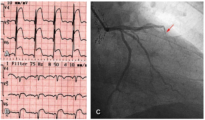
Figure 1A. ST segment elevation with increased magnitude (9 mm) in V4-V6 which reflects the myocardium at risk. Figure 1B. Transformation of the myocardium at risk in established infarction, Q wave V4-V5 aspect with the resolution of the R wave. Figure 1C. Occlusion of anterior descending artery in the medial segment (arrow). Personal case.
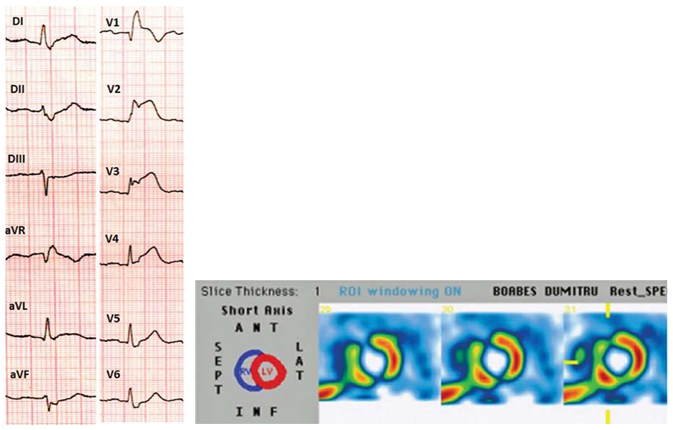
Figure 2A. The magnitude of the ST segment elevation higher than the R wave amplitude in V2, V3 leads. Figure 2B. The scintigraphy demonstrates the occurance of myocardial infarction with antero-septal location. Personal case.
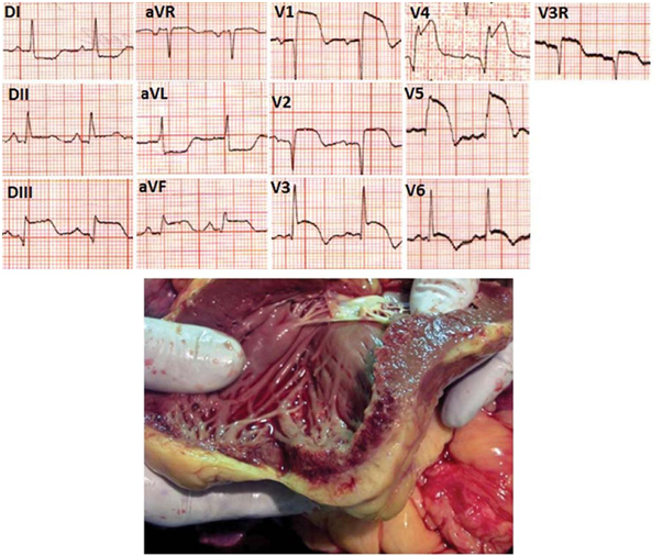
Figure 3A. Antero-lateral, inferior and right ventricle myocardial infarction. Tombstone ST segment elevation in V4,V5 leads (ST nadir > R wave amplitude). Figure 3B. Anatomopathological examination of the patients mentioned above. There can be remarked hemorrhagic necrosis lesions of the inferior, ante-rior and right ventricle walls. Personal case.
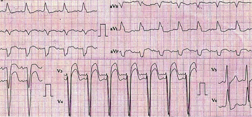
Figure 4. Left bundle branch block. ST segment elevation > 5 mm in V3, V4 leads suggesting associated myocardial infarction. Personal case.
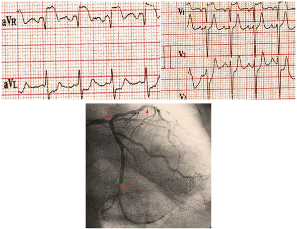
Figure 5A. ST segment elevation in aVR and V1 leads with ST depression in aVL, V3 leads, aspect that suggests an acute myocardial infarction determined by left coronary artery occlusion. Figure 5B. Signifi cant stenosis of the left coronary artery, anterior descending artery and circumflex artery (arrows) – the same pacient described above. Personal case.
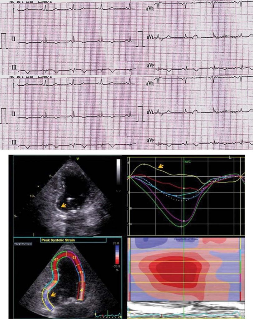
Figure 6A. Necrosis Q waves noticeable in DII, DIII, aVF leads associated with ST segment elevations and negative T waves. This aspect is suggestiv for inferior wall aneurysm of the left ventricle. Figure 6B. It can be distinguished the aneurysmal aspect of the inferior wall (thin arrow) in the patient above. The strain value of the inferior wall of left ventricle during the systolic period is positive suggesting an aneurysm (big arrow). Personal case.
CONCLUSIONS
- Electrocardiographic assessment has to be initial and it is essential in diagnosis of acute coronary occlusion, at the same time underlying the thera-peutic approach.
- Evaluation regarding magnitude and extension of the ST segment represents a total different assay in comparison to radionuclidic techniques or nu-clear magnetic resonance used for the evaluation of the myocardium at risk of necrosis.
- Information about morphopathologic features of ST segment elevation in case of acute myocardial infarction provides additional details to the ana-tomic and metabolic changes.
- In my opinion, the presence of a „momentum” which identifies a major amplitude of ST segment elevation constiutes, however, a reference point in establishing the severity of the myocardial in-jury and in adopting a quick decision regarding proper therapeutic approach.
References
1. Thygesen C, Alpert JS, Jaffe AS et al. Fourth Universal Definition of Myocardial Infarction. Circulation 2018;138:e618-e651.
2. Timothy F, Christian MD, Raymond J et al. Estimates of Myocardi-um at risk and Collateral Flow in Acute Myocardial Infarction Using Electrocardiographic Indexes with Comparison to Radionuclide and Angiographic Measures. J Am Coll Cardiol 1995;26:388-393.
3. Holland RP, Arnsdorf MF. Solid Angle Theory and the Electrocardio-gram: Physiologic and Quantitative Interpretations. Prog Cardiovasc Dis 1977;19(6):431-57.
4. Aldrich H, Wagner N, Boswich J et al. Use of Initial ST-Segment De-viation for Prediction of Final Electrographic Size of Acute Myocar-dial Infarcts. Am J Cardiol 1988;61(10):749-53.
5. Idelkar RE, Wagner GS, Ruth WK et al. Estimation of a QRS Scor-ing System for Estimating Myocardial Infarct Size. Correlation with Quantitative Anatomic Findings for Anterior Infarcts. Am J Cardiol 1982;49:1604-14.
6. Shaimaa, Mohamed A, Heba A. Electrocardiographic Predic-tor of Final Infarct Size by Selvester QRS Score in Correlation to Rest Tc99M Sestamibi SPECT afer Primary PCI. Med J Cairo Univ 2015;83(1):499-506.
7. Yang ZK, Shen Y, Hu J et al. Impact of Coronary Collateral Circu-lation on Angiographic In-stent Restenosis in Patients with Stable Coronary Artery Disease and Chronic Total Occlusion. In J Cardiol 2017;15:247:26.
8. Mauri F, Gasparini M, Barbonaglia L et al. Prognostic Significance of the Extent of Myocardial Injury in Acute Myocardial Infarction Treat-ed by Streptokinase (The GISSI trial). Am J Cardiol 1989;63:1291-95.
9. Lemkes K, Janssens NG, der Hoeven NW et al. Timing of Revascu-larization in Patients with Transient ST-Segment Elevation Myocar-dial Infarction: a Randomized Clinical Trial, Eur Heart J 2019;40:283-291.
10. Balci B. Tombstoning ST-Elevation Myocardial Infarction. Current Cardiology Reviews 2009;5:273-278.
11. Ibanez B, James S, Agewall S et al. ESC Guidelines for the Manage-ment of Acute Myocardial Infarction in Patients Presenting with ST-Segment Elevation: The Task Force for the Management of Acute Myocardial Infarction in Patients Presenting with ST-Segment El-evation of the European Society of Cardiology 2017. Eur Heart J 2018;39(2):119-177.
12. Ortiz TJ, Meyers NS, Davidson SC et al. Impact of Residual Flow on the Reduction in Infarct Transmurality and Preservation of Myocar-dial Salvage Following Reperfused ST-Segment Elevation Myocardial Infarction, Circulation 2018;114:343.
13. Willems JL, Willems RJ, Willems GM et al. European Cooperative Study Group for Recombinant Tissue Type Plasminogen Activator Significance of Initial ST-Segment elevation and Depression for the Management of Thrombolytic Therapy in Acute Myocardial Infarc-tion. Circulation 1990;82:1147-58.
14. Klein LR, Shroff GR, Beeman W et al. Electrocardiographic Criteria to Differentiate Acute Anterior ST-elevation Myocardial Infarction from Left Ventricular Aneurysm. Emerg Med 2015;33(11);1707-08.
15. Hasan A, Muzamil M, Aftab A et al. Correlation of Type of ST Seg-ment Elevation in Acute Anterior Wall Myocardial Infarction on Electrocardiogram with Left Ventricular Ejection Function. J Interv Gen Cardiol 2018;2(2):116. -
 This work is licensed under a