Marin Postu1, Lucian Predescu1
1 “Prof. Dr. C.C. Iliescu” Emergency Institute for Cardiovascular Diseases, Bucharest, Romania
We present the case of a 66-year-old male patient admitted in our institute for aggravated angina. His cardiovascular risk factors were dyslipidemia and stage III hypertension. The resting electrocardiogram was unremarkable, and the electrocardiographic stress test was positive for ischemia. The transthoracic echocardiography showed a normal left ventricular ejection fraction and mild mitral regurgitation. Coronary angiogram revealed a mild distal left main stenosis, left anterior descending artery with a moderate stenosis at the ostium and a tight stenosis in the proximal segment, left circumflex artery, ramus intermedius artery and right coronary artery with atheromatous plaques (Figure 1). The heart team decided to revascularize the patient by percutaneous coronary intervention. After predilatation of the lesion on proximal left anterior descending artery, a 3.0-3.5×27 mm self apposing stent was implanted from left main toward left anterior descending artery. The stent was postdilated with a 3.5mm noncompliant balloon and the stent struts were opened toward left circumflex artery with a 2.5 mm semicompliant balloon. A good final angiographic result was obtained (Figure 2). Intravascular ultrasound showed a severe deformation of proximal part of the stent with a severe malapposition in the left main most likely because the guiding catheter came under the stent when we removed the sheath and balloon of the stent (Figure 3). Post dilatation of the stent from the left main was done with a 4.0 mm noncompliant balloon at very high pressure. Intravascular ultrasound was repeated that showed the correction of stent deformation and malapposition with an optimal minimum instent area (Figure 4).
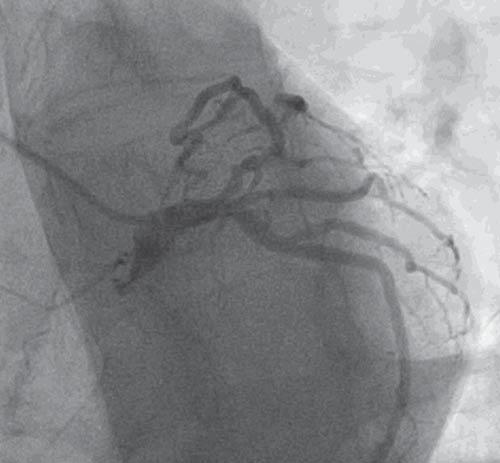
Figure 1. Coronary angiogram – left coronary artery – mild distal left main stenosis, left anterior descending artery with a moderate stenosis at the ostium and a tight stenosis in the proximal segment, left circumflex artery, ramus intermedius artery with atheromatous plaques.
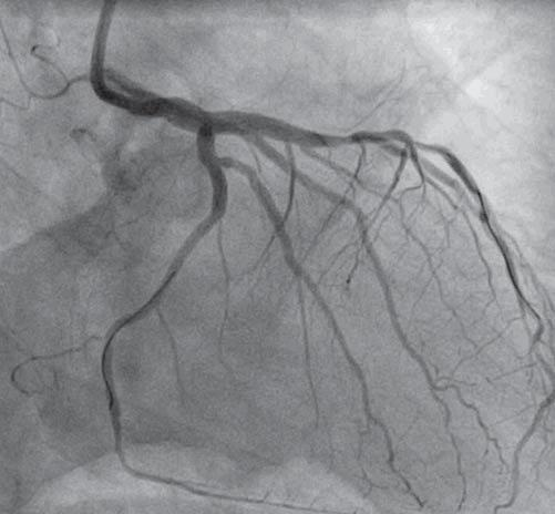
Figure 2. Coronary angiogram – left coronary artery – final angiographic result after percutaneous coronary intervention.
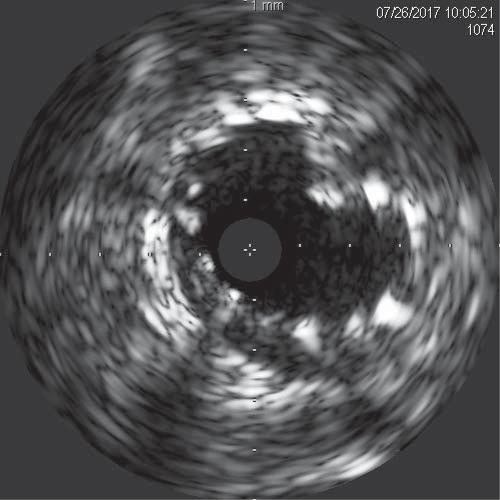
Figure 3. Intravascular ultrasound – a severe deformation of proximal part of the stent with a severe malapposition in the left main.
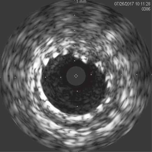
Figure 4. Intravascular ultrasound – correction of stent deformation and malapposition after postdilatation with an optimal minimum instent area.
 This work is licensed under a