Laura Tapoi2, Radu A. Sascau1,2, Cristina Cristea4, Viorel Scripcariu1,3, Raluca Chistol1,2, Alexandra Clement2, Stefan Boca2, Larisa Anghel1,2, Cristian Statescu1,2
1 „Grigore T. Popa” University of Medicine and Pharmacy, Iasi, Romania
2 „Prof. Dr. George I.M. Georgescu” Institute of Cardiovascular Diseases, Iasi, Romania
3 Ist Clinic of Oncological Surgery, Regional Institute of Oncology, Iasi, Romania
4 Clinic of Endocrinology, „Sf. Spiridon” University Hospital, Iasi, Romania
Abstract: Objective – Arterial hypertension is an important cardiovascular risk factor with destructive effects on the cardio-renal axis. Approximately 10% of the hypertensive population suffers from the secondary form of this pathology. Methods – We hereby present the case of a 47-year-old patient who was addressed to our clinic because of persistent high blood pressure values, despite medication compliance. Results – Laboratory fi ndings revealed elevated creatinine and hypo-kalaemia. Transthoracic echocardiography revealed left ventricular hypertrophy, diastolic dysfunction and subclinical systolic dysfunction. The renal angiogram was normal. The aldosterone: renin ratio was elevated. The tomographic computer exam revealed the presence of two micronodules in the right adrenal gland. The diagnosis of primary hyperaldosteronism was es-tablished. After the association of an antialdosteronic agent, a better control of the tensional values was obtained. The pati-ent was referred for the surgical treatment of the lesion. Conclusions – Primary hyperaldosteronism accounts for 5-10% of resistant hypertension cases, and unilateral adrenal adenomas are the second most common cause after bilateral idiopathic hyperplasia. When untreated, it is associated with an increased rate of arrhythmias, coronary artery disease, heart failure, stroke, proteinuria and renal dysfunction. The gold standard for the treatment of unilateral adenomas is surgical resection. Keywords: secondary hypertension, hyperaldosteronism.
INTRODUCTION
Arterial hypertension is an important cardiovascular risk factor with destructive effects on the cardio-renal axis. Approximately 10% of the hypertensive populati-on suffers from the secondary form of this pathology1. The identifi cation of the secondary causes of hypertension is important, as they may be curable. Further-more, when left undiagnosed, secondary hypertensi-on can lead to cardiovascular and renal complicati-ons, with an increased mortality and burden on the healthcare system.
CASE PRESENTATION
A 47-year-old patient presented with persistent high blood pressure values, in spite of good compliance with maximal medical therapy. His medical history re-vealed a recent ischemic stroke with right-sided he-miparesis and arterial hypertension diagnosed 7 years ago.
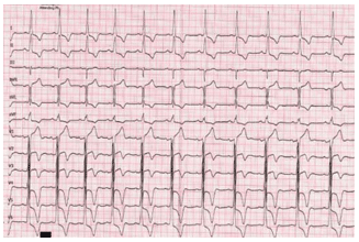
At admission, the clinical examination revealed no pathological signs, except for a motor deficit on the upper and lower right limbs. The blood pressure was 210/110 mmHg and the heart rate was 65 beats per minute. Resting ECG showed sinus rhythm and left ventricular hypertrophy (LV) with left ventricular stra-in (Figure 1).
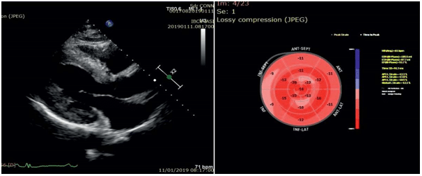
Laboratory findings at admission revealed hypokale-mia (K= 3.2 mmol/L) and renal impairment with an es-timated glomerular filtration rate (eGFR) of 51.3 mL/ min/1.73 m2. Transthoracic echocardiography showed left ventricular hypertrophy, with LV ejection fraction within normal limits, with longitudinal systolic LV dys-function and LV diastolic dysfunction with increased filling pressures (Figure 2). The 24-hour blood pre-ssure (BP) monitoring revealed a mean BP of 187/102 mmHg with a maximum BP of 217 mmHg, a minimum systolic BP of 138 mmHg and a non-dipper profile.
When discussing the causes of resistant hyperten-sion, after excluding the pseudo-resistant situations as poor adherence to medical therapy and white-coat phenomenon, the following forms of secondary hyper-tension were taken into consideration: renovascular disease, renal parenchymal hypertension and endocri-ne causes. Obesity, excessive alcohol consumption, high sodium intake and obstructive sleep apnoea were too considered, and for the latter the patient was di-rected to polysomnography at discharge.
For establishing the existence of a renovascular di-sease, an abdominal ultrasound with colour Doppler of the renal arteries was performed. The renal arte-ries could not be evaluated because of poor acous-tic window (abdominal obesity), but the abdominal ultrasound showed an asymmetry of approximately 15 mm between the kidneys: right kidney 85/40 mm, left kidney 102/56 mm. Therefore, a renal angiogra-phy was performed, which excluded the presence of significant stenosis on the renal arteries. The patient had no history of urinary tract infections, the urinalysis was normal and there were no signs of vesicoureteral refl ux or other causes of urinary tract obstruction, thus a chronic pyelonephritis was improbable. The la-boratory tests were negative for phaeochromocyto-ma, Cushing’s syndrome, thyroid disease and hyper-parathyroidism. Plasma aldosterone and renin, and aldosterone: renin ratio values were as follows: renin =14.4 ng/L, aldosterone 797 ng/L, aldosterone: renin ratio=55.3. The adrenal computed tomography (CT) showed two small nodules on the right adrenal gland (Figure 3).
The abovementioned results were consistent with the diagnosis of primary hyperaldosteronism. After the inclusion of an antialdosteronic drug in high dose in the antihypertensive regimen, the 24-hour BP mo-nitoring revealed a mean BP of 152/91 mmHg, with a maximum systolic BP of 179 mmHg and a minimum systolic BP of 123 mmHg. The patient was referred to surgery for the laparoscopic excision of the right adrenal gland. The diagnosis of adrenal adenoma was confirmed by the anatomopathological examination. After surgery, an optimal control of the blood pre-ssure values was obtained under treatment with beta-blocker, alpha blocker and calcium channel blocker.

Figure 1. Resting ECG: sinus rhythm 60 beats per minute, left ventricular hypertrophy with left ventricular strain.

Figure 2. Transthoracic echocardiography, left: parasternal long axis view, LV hypertrophy; right: speckle tracking-longitudinal LV systolic dysfunction.
DISCUSSION
In 1955, J.W.Conn described for the first time the pri-mary aldosteronism (PA) as a syndrome characterised by „the presence in the urine of excessive amounts of a sodium-retaining corticoid, severe hypokaelmia, hypernatremia, alkalosis, and a renal tubular defect in the reabsorption of water”. PA can also present with normokalemia, in which case the hypertension can be misdiagnosed as essential hypertension (EH)2. Different studies have reported a prevalence of PA about 5-10% of hypertensive patients3. The prevalence of PA increases with the severity of hypertension and patients with PA display more frequently target or-gan damage (LV hypertrophy, microalbuminuria) and cardiovascular events, with an increased mortality4,5.
Moreover, in a study which included 553 patients, the prevalence of cardiovascular events was signifi cantly higher in PA patients with hypokalemia6. When com-pared to patients with EH, PA patients have greater deterioration of LV diastolic function and a higher pre-valence of eccentric hypertrophy7. Furthermore, PA patients have greater subclinical systolic dysfunction than EH patients8.
For the diagnosis of PA, the European Society of En-docrinology recommends screening in high risk popula-tion, by measuring the plasma aldosterone and renin values and aldosterone: renin ratio. The next step is represented by confirmatory testing (saline loading, fludrocortisone or captopril challenge) which is con-sidered mandatory, with an exception represented by PA cases presenting with spontaneous hypokalemia and a plasmatic aldosterone >200ng/L. CT scanning or magnetic resonance imaging are recommended for the subtype differentiation of PA, but the adrenal vein sampling is recommended in candidates for surgery, as it has greater specificity in differentiating unilateral from bilateral PA9. However, the SPARTACUS trial showed that treating PA patients based on CT scan-ning was non-inferior in terms of antihypertensive treatment intensity and blood pressure control and even superior in terms of associated financial costs10.
The gold standard for the treatment of unilateral PA is represented by adrenalectomy (9), but unfortu-nately not all the patients are cured after the surgical intervention. A recent meta-analysis which included 37.763 patients reported a mean hypertension cure rate after unilateral adrenalectomy in PA patients of 50.6%11.
The presented case illustrates the aspects discussed above and is particular because of the late diagnosis of PA, after 7 years of evolving uncontrolled hypertensi-on, in a patient with established organ damage (stro-ke, renal impairment). The chronic exposure to high aldosterone levels leads to myocardial and vascular fibrosis, endothelial dysfunction and microangiopathy and can explain the necessity of continuing the anti-hypertensive therapy post adrenalectomy.
CONCLUSIONS
Although potentially curable, PA continues to be an underdiagnosed disease. Its diagnosis is important and screening in high risk populations should be perfor-med, because when PA remains undiagnosed it associ-ates an increased rate of arrhythmias, coronary artery disease, heart failure, stroke, proteinuria and renal dysfunction. The gold standard for the treatment of unilateral adenomas is represented by surgical resec-tion.
Conflict of interest: none declared.
References
1. Puar TH, Mok Y, Debajyoti R, Khoo J, How CH, Ng AK. Secondary hypertension in adults. Singapore medical journal. 2016;57(5):228-32.
2. Conn JW. Presidential address. I. Painting background. II. Primary al-dosteronism, a new clinical syndrome. The Journal of laboratory and clinical medicine. 1955;45(1):3-17.
3. Er LK, Wu VC. Call for screening for primary aldosteronism: an underdiagnosed and treatable disease. Journal of thoracic disease. 2018;10(2):557-9.
4. Monticone S, Burrello J, Tizzani D, Bertello C, Viola A, Buffolo F, et al. Prevalence and Clinical Manifestations of Primary Aldosteronism Encountered in Primary Care Practice. Journal of the American Col-lege of Cardiology. 2017;69(14):1811-20.
5. Reincke M, Fischer E, Gerum S, Merkle K, Schulz S, Pallauf A, et al. Observational study mortality in treated primary aldosteronism: the
German Conn’s registry. Hypertension (Dallas, Tex: 1979). 2012; 60(3):618-24.
6. Born-Frontsberg E, Reincke M, Rump LC, Hahner S, Diederich S, Lorenz R, et al. Cardiovascular and cerebrovascular comorbidities of hypokalemic and normokalemic primary aldosteronism: results of the German Conn’s Registry. The Journal of clinical endocrinology and metabolism. 2009;94(4):1125-30.
7. Yang Y, Zhu LM, Xu JZ, Tang XF, Gao PJ. Comparison of left ventric-ular structure and function in primary aldosteronism and essential hypertension by echocardiography. Hypertension research : official journal of the Japanese Society of Hypertension. 2017;40(3):243-50.
8. Chen ZW, Huang KC, Lee JK, Lin LC, Chen CW, Chang YY, et al. Aldosterone induces left ventricular subclinical systolic dysfunction: a strain imaging study. Journal of hypertension. 2018;36(2):353-60.
9. Williams TA, Reincke M. MANAGEMENT OF ENDOCRINE DIS-EASE: Diagnosis and management of primary aldosteronism: the En-docrine Society guideline 2016 revisited. European journal of endo-crinology. 2018;179(1):R19-r29.
10. Dekkers T, Prejbisz A, Kool LJS, Groenewoud H, Velema M, Spiering W, et al. Adrenal vein sampling versus CT scan to determine treat-ment in primary aldosteronism: an outcome-based randomised diag-nostic trial. The lancet Diabetes & endocrinology. 2016;4(9):739-46.
11. Zhou Y, Zhang M, Ke S, Liu L. Hypertension outcomes of adrenal-ectomy in patients with primary aldosteronism: a systematic review and meta-analysis. BMC endocrine disorders. 2017;17(1):61.
 This work is licensed under a