Download PDF
https://doi.org/10.47803/rjc.2020.30.2.257
Georgiana Poparlan1*, Mirela Mihaela Mihalcia1*, Ovidiu Mitu1,2, Cristina Tarniceriu2*, Cristian Statescu2,3, Viviana Onofrei1,2, Razan Al Namat2, Ana Maria Buburuz1,2, Irina Iuliana Costache1,2, Antoniu Octavian Petris1,2
1 Department of Cardiology, „Sf. Spiridon” Clinical Emergency Hospital, Iasi, Romania
2 „Grigore T. Popa” University of Medicine and Pharmacy, Iasi, Romania
3 „Prof. G.I.M. Georgescu” Institute of Cardiovascular Diseases, Iasi, Romania
* equal contribution
Abstract: Treatment of patients with immune thrombocytopenic purpura (ITP) associated with recurrent venous and arterial thrombosis can represent a major challenge. We present the rare case of a 56-year-old female who was first diagnosed with severe ITP at the age of 36. She required corticosteroid therapy and splenectomy in evolution. However, in the last three years she had several episodes of recurrent venous thromboembolism for which she required different anticoagulant therapies despite severe thrombocythemia. The patient also developed acute myocardial infarction treated by primary percutaneous coronary intervention that was complicated with acute intrastent thrombosis. Thus, maximal antiplatelet therapy was mandatory. For ITP, the patient received intravenous steroids, platelet transfusion as well as eltrombopag. Moreover, the patient also suffered from a haemorrhagic uterine fibroma that required surgery. Thus, a close multidisciplinary approach was needed for the successful treatment of this patient.
Keywords: thrombocytopenia; thromboembolism; myocardial infarction; thrombosis; immune purpura; anticoagulant.
Rezumat: Tratamentul pacienţilor cu purpură trombocitopenică imună (PTI) asociată cu tromboză arterială şi venoasă recurentă poate reprezenta o importantă provocare. Prezentăm cazul unei paciente de 56 ani, diagnosticată de la vârsta de 36 ani cu PTI care a necesitat terapie cu corticosteroizi şi splenectomie în evoluţie. Totuşi, în ultimii trei ani, pacienta a prezentat multiple episoade de tromboembolism venos recurent care a necesitat diverse terapii anticoagulante în ciuda trombocitopeniei severe. Ulterior, pacienta a dezvoltat infarct miocardic acut tratat prin angioplastie percutană coronariană primară, complicat cu tromboză acută intrastent. Prin urmare, terapia antiplachetară maximală a fost obligatorie. Pentru PTI, pacienta a primit steroizi intravenos, transfuzie plachetară, precum şi eltrombopag. Mai mult, pacienta a prezentat un fi brom uterin hemoragic care a necesitat intervenţie chirurgicală în ciuda trombocitopeniei severe. Prin urmare, o abordare multidisciplinară a fost necesară pentru terapia de succes a acestei paciente.
Cuvinte cheie: trombocitopenie; tromboembolism; infarct miocardic; tromboză; purpura imună; anticoagulare.
INTRODUCTION
Immune thrombocytopenic purpura (ITP) is a rare autoimmune disease characterized by autoantibody-mediated platelet destruction along with suppression of their production1. ITP can be asymptomatic or can manifest with bleeding due to the low platelet count2. However, paradoxically, it is associated with thrombotic events as well3. There are several data regarding the risk of thrombosis in ITP patients4. In a meta-analysis of observational studies, the adjusted relative risk for arterial and venous thromboembolism in ITP patients was 1.5 and 1.9, respectively5. However, the-re are currently no guidelines regarding anticoagulati-on in ITP patients despite the increased risk of throm-bosis. Moreover, the appropriate use of anticoagulant therapy in ITP patients who develop thrombosis re-lapse together with thrombocytopenia has not been comprehensively studied.
Thus, we present the case of a patient with ITP who developed multiple recurrences of deep vein thrombosis (DVT) and pulmonary thromboembolism (PE) despite anticoagulant therapy, while being in thrombocytopenic crisis.
CASE REPORT
We report the case of a 56-year-old female patient, with history of ITP from the age of 36. She was initially placed on chronic corticosteroid therapy. She eventually required splenectomy due to refractoriness to medical therapy and multiple relapses during steroid tapers. During corticosteroid treatment, the patient developed several complications such as diabetes mellitus, arterial hypertension, osteoporosis and glaucoma. At the age of 53 years and 3 months, two weeks after her splenectomy, the patient was referred to the Cardiology Clinic for painful left lower extremity swelling with high DVT suspicion. Heart, lung and abdominal exams were unremarkable. There were no cutaneous or mucosal petechie or purpura. Blood tests showed haemoglobin (Hb) 9.8 g/dl, white blood cell count 16.000/μL and platelet count (PLT) 104.000/μL, normal prothrombin time (PT). Electrocardiogram and transthoracic echocardiogram (TTE) were unremarkable. DVT was confirmed by venous Doppler ultrasound. This revealed thrombus in the left superficial femoral, popliteal and proximal calf ve-ins. During hospitalization, the patient received 5 days of intravenous unfractioned heparin (UFH), closely aPTT-adjusted. She was bridged to oral anticoagulati-on with vitamin K antagonist (VKA). Despite medication compliance, she had recurrence of the left lower extremity DVT one month later. She was again placed on intravenous UFH and then transitioned to a direct oral anticoagulant (DOAC) – dabigatran 150 mg b.i.d.
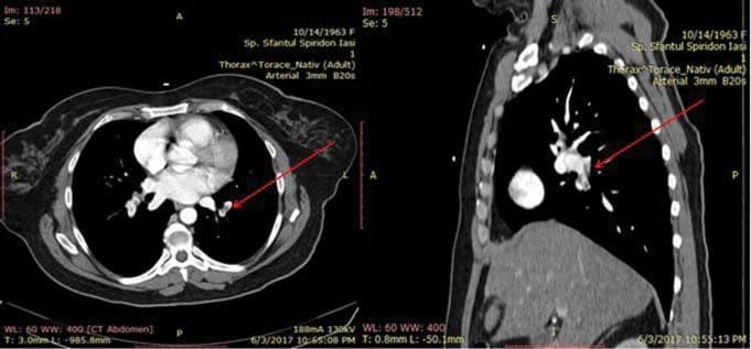
One year later, she was hospitalized at the Gynecology for a bleeding uterine fibroma. She required emergent surgical intervention and interruption of her oral anticoagulation. She developed sudden on set resting dyspnea and was readmitted in the Cardiology Clinic. She was afebrile, heart rate 100 bpm, blood pressure 100/70 mmHg, oxygen saturation 90% in room air. Her complete blood count (CBC) showed: Hb 6.4 g/dl, WBC 25.000/μL and PLT 3.000/ μL (day 0). Among cardiac markers, D-dimers were very elevated and PE was suspected. The pulmonary computed-tomography (CT) angiography confirmed the diagnosis. It revealed newly developed bilateral PE, predominantly in the left pulmonary artery (Figure 1). Abdominal ultrasound revealed an expanding, well delimited, inhomogeneous, right parauterine structure with maximum diameter of 150 mm and with positive Doppler signal. Workup for lupus including lupus anti-coagulant, elevated immunoglobulin G, anticardiolipin antibody titres were all negative. By further analysis, hyperhmocysteinemia or other types of thrombophilia and coagulation factor deficit have been excluded.
Under the advice of a haematologist, the patient received PLT transfusion and steroid therapy. On day 3 of hospitalization, PLT increased to 49.000/μL and intravenous UFH was initiated. On day 6, the patient had no menorrhagia or other bleeding and was swit-ched to weight-adjusted low-molecular-weight heparin (LMWH) – enoxaparin. The Hb increased to 10.4 g/dl, but thrombocytopenia relapsed without active bleeding. On day 10 of hospitalization, the PLT count decreased to 3.000/μL, so intravenous steroids were administered. Subsequently, to maintain PLT count in a safe range, thrombopoietin-receptor agonist (TPO-RA) eltrombopag was initiated, with good response. She remained hemodynamically stable and was dis-charged after 18 days of hospitalization. On discharge, her Hb was 11.9 g/dl, PLT of 35.000/μL. The choice of anticoagulant treatment at discharge was a challenge, balancing the thrombotic – haemorrhagic risk, however dabigatran 150 mg b.i.d. was maintained.
Two months later (54 years and 8 months), the patient presented to the emergency room with 3-day history of continuous typical angina. Her electrocar-diogram and elevated troponin level confirmed an acute anterolateral ST segment elevation myocardial infarction (STEMI). Laboratory blood tests showed Hb 12.5 g/dl, PLT count 19.000/μL and normal PT. Prior to coronary angiography, intravenous dexamethasone and platelet transfusion were administrated, as advised by the haematology department, due to temporary lack of immunoglobulins. She underwent emergent coronary angiogram that revealed acute thrombotic occlusion of the first segment of the left anterior descending artery (LAD). After thrombus aspiration, a drug eluted stent was successfully deployed in the LAD. 2 days after PCI, she had sudden recurrence of chest pain. Urgent coronarography revealed acute intrastent thrombosis that benefi ted from thromb aspiration. She was discharged with post STEMI treatment that included triple therapy with aspirin, clopidogrel and dabigatran 150 mg b.i.d, as well as eltrombopag for the ITP.
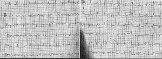
At the age of 56 years and 2 months, one year and a half from her STEMI, the patient was admitted once again in the Cardiology Clinic with suspected DVT: right lower extremity oedema and pain. She had normal pulse rate 80 bpm, respiratory rate 16/min, blood pressure 135/80 mmHg and oxygen saturation 97% in room air. Venous Doppler ultrasound revealed occlusive thrombus of the right superficial femoral and popliteal veins. The electrocardiogram recorded sinus rhythm, 80 bpm, QRS axis -20 degrees, pre-existent complete right bundle branch block (RBBB) and sequela of old anterior myocardial infarction (Figure 2). Laboratory tests showed Hb 11 g/dl, WBC 14.400/ μL and PLT count 13.000/μL, aPTT 29 s, normal renal and liver function. The transthoracic echocardiogram revealed dilated left ventricle with moderate systolic dysfunction (ejection fraction 40%) with akinesia of the left ventricular apex and interventricular septum and hypokinesia of the anterior wall, moderate functional mitral regurgitation.
During hospitalization, the patient received intravenous UFH, switched afterwards to fondaparinux 7.5 mg once daily. Her aspirin was continued. After 3 months of treatment, follow-up venous Doppler ultra-sound showed improvement of her DVT. However, she rapidly developed menorrhagia with Hb drop to 7 g/dl and PLT of 4.000/μL. Thus, the dose of fondapa-rinux was reduced to 2.5 mg once daily and the anti-platelet therapy was stopped. Uterine fibroma surgery was successfully performed under optimal conditions. Afterwards, the patient has been closely followed up in the cardiology and haematology departments. To date, she has not had any further complications. Table 1 presents schematically the pathological background of the patient.
The particularities of this case are represented by the appearance of successive thrombosis recurrences even after the initiation of the anticoagulant therapy in a patient with ITP exacerbation. On the background of thrombocytopenia, subsequent occurrence of an acute coronary syndrome which required stent im-plantation and complicated with acute onset of intrastent thrombosis is also very rare. The coexistence of uterine fibroma that caused major bleeding and required surgery represented an obstacle in the continuous administration of the anticoagulant and antiplatelet therapy that could have threatened the patient’s prognosis.

Figure 1. Pulmonary CT angiography: filling defect in both pulmonary arteries, predominantly on the left side.

Figure 2. Electrocardiography: sinus rhythm, 80 bpm, complete RBBB, chronic anterior MI sequela.

DISCUSSION
Although the greatest danger of ITP is life-threatening bleeding during severe thrombocytopenia2, this disea-se has, paradoxically, prothrombotic effects as well3. Thrombocytopenia does not protect ITP patients against thromboembolic disease. Arterial and venous thromboembolism (VTE) events occur in ITP patients as they occur in the normal population6 and the risk seems to be higher with lower platelet counts7-9. Some studies showed that the patients with primary immune thrombocytopenia are at increased risk for venous thromboembolic events compared with patients without primary immune thrombocytopenia and knowledge of an elevated risk of venous thrombosis among adult patients with primary immune thrombo-cytopenia may possibly support increased utilization of thromboprophylactic treatment in patients at lower risk of haemorrhage3. We considered that some important factors are implicated in pathophysiology of the thrombotic events in this clinical setting: ITP itself, ITP treatment and other comorbidities and diseases.
May ITP itself induce thrombotic events? The pathophysiology showed that ITP can be considered an inflammatory disease with elevated proinflamma-tory cytokines (IL-6, IL- 21) and decreased the level of the Treg cells. The presenting antigen cells, B and T lymphocytes interact between them and produce an inflammatory status. The inflammatory state can interfere with haemostasis leading to a hypercoagulation. An important role in thrombosis is attributed to the microparticles (MPs) that are small vesicles that result from blebbing of the cellular membrane during acti-vation or apoptosis processes10. The MPs are derived from a variety of cells type including the platelets, mo-nocytes and endothelial cells11. Possible explanations for the tendency of ITP patients to have thromboses may be related to increased proportion of platelet microparticles which are the result of autoantibody – induced platelet fragmentation that cause platelet activation12,13. The platelet microparticles (PMPs) are vesicles derived from platelet membranes that arise in association with platelet activation and other unknown ca-uses. Elevated PMPs have been observed in idiopathic thrombocytopenic purpura (ITP) and concluded that PMPs play an important role in haemostasis, and that high concentrations of hemostatically active PMP can be thrombogenic12. The presence of anti-platelet autoantibodies increases the risk of thrombotic events perhaps due to procoagulant microparticles released by activated platelets or associated predispositions14. Monocyte MPs are an important sources of circulating tissue factor for arterial thrombus formation10. In response to a variety of agonists, including cytokines, the endothelial cells express the tissue factor which is the major cellular activator of the clotting cascade. MPs released from endothelial cells and their interactions with leukocytes are likely to play a central role on thrombogenesis15.
Treatment for ITP may also be associated with thrombotic complications. Corticosteroids can play a role in the formation of a hypercoagulable state16-19. Intravenous immunoglobulin therapy (IVIG) contributes to the increased risk of thrombosis via increased plasma viscosity, platelet aggregation, complement activation, as described in previous studies20-21. Moreover, newly introduced thrombopoietin-receptor agonists, such as eltrombopag and romiplostim, seem to increase thrombotic risk, although the mechanisms remain unsettled4,22. Moreover, splenectomy increases the risk of thrombotic events23.
Reports described that ITP patients with positive antiphospholipid antibodies (lupus anticoagulant and anticardiolipin antibodies), have a significantly increased risk for thrombosis24-26 but in our case these anti-bodies were negative. However, age, personal risk factors may put some ITP patients at a particularly higher risk of thrombosis.
The presence of thrombotic events in patients with ITP represents a challenge in terms of therapeutic management. Both VTE and myocardial infarction may occur when platelets are low. In the case of occurrence, a careful balance between usual anticoagulation and antiplatelet therapy on one hand and efforts to raise platelet count on the other hand is needed. There is few literature data available for the management of thrombotic events in ITP patients. Matzdorff et al. propose a therapeutical algorithm for patients with thrombocytopenia and high bleeding risk. They suggest the administration of anticoagulant up to a platelet count of more than 30.000/μL, only if no li-fe-threatening or need of transfusion bleedings occur. In the first 48 hours, it is preferable to start treatment with UFH. If no bleeding occurs, the transition to LMWH is suitable27. But there are no data when to start anticoagulant treatment.
DOAC are widely used for treatment of acute PE and DVT28, but their safety and efficacy in patients with ITP have not been thoroughly evaluated. They are more accessible than AVK because there is no need for treatment monitoring. Dabigatran acts by inhibiting thrombin, but it is possible that isolated inhibition of thrombin may not be enough when prothrombin status is intense. We have chosen dabigatran due to its reversibly binding to the active site on the throm-bin molecule, preventing thrombin-mediated activation of coagulation factors. Moreover, there are no studies to show whether dabigatran is less effective than other DOAC in ITP.
In our reported case, the patient had multiple episodes of VTE concomitantly with severe thrombo-cytopenia. Estimating the risk of bleeding and the risk of VTE progression is useful before starting the therapy. Standardized therapy was recommended while patient was in remission. PE occurred in the context of discontinuation of anticoagulant therapy due to the development of menorrhagia associated with platelet count of 3.000/μL. Thus, it was considered for pretreatment with platelet support and steroids while postponing the anticoagulant therapy. As the risk of bleeding is present, the option remains the usage of UFH while fondaparinux has been proposed as an alternative to UFH. Unlike UFH and LMWH, fondaparinux is a pure antithrombin III-dependent factor Xa inhibitor which has no effect on platelet function. This may potentially decrease the risk of serious bleeding in thrombocytopenic patients29-31. In the case of anticoagulant medication contraindications, it is an option to use a filter on the inferior vena cava.
In the present case, the patient developed acute coronary syndrome for which she received a drug eluted stent because of the lesion location that predisposed to a high risk of restenosis. Unfortunately, the patient developed acute intrastent thrombosis. Thrombopoietin analogues have been known to be prothrombotic and an incident of stent thrombosis has been reported in a patient on romiplostim32.
Thus, multidisciplinary approach (cardiologist, haematologist) is required for the proper management of certain medication interactions. In all the reviewed cases, the antiplatelet therapy was safe and well tolerated even for long periods of time and was only discontinued during bleeding episodes. Among the ca-ses found in the literature, 14 patients were discharged with aspirin, 15 patients received clopidogrel, 2 patients received abciximab, while 3 patients did not receive antiplatelets32-36. During antiplatelet treatment, only one patient presented petechiae and epistaxis that required discontinuing clopidogrel37. There is a need for additional research into the pathophysiology of relapsed thrombotic events in the context of ITP and its therapeutic influence.
CONCLUSION
There is limited data that guide therapeutic management of ITP patients with arterial and venous thromboembolism. It is important to carefully evaluate the risk of bleeding vs. the risk of thrombosis before initiating antithrombotic therapy. UFH should be considered the first option in stabilized haematological status. As for the thrombocytopenic crisis or active bleeding, proper management of ITP with steroids, IVIG or platelet transfusion should be considered. Dual antiplatelet therapy associated with anticoagulant increases the risk of bleeding in ITP patients, thus further studies are needed to better define a personalized antithrombotic approach.
Conflict of interest: none declared.
References
1. Nugent D, McMillan R, Nichol JL, et al. Pathogenesis of chronic im-mune thrombocytopenia: increased platelet destruction and/or de-creased platelet production. Br J Haematol. 2009;146(6):585-596.
2. Cohen YC, Djulbegovic B, Shamai-Lubovitz O, et al. The bleeding risk and natural history of idiopathic thrombocytopenic purpura in patients with persistent low platelet counts. Arch Intern Med. 2000; 160(11):1630-1638.
3. Sarpatwari A, Bennett D, Logie JW, et al. Thromboembolic events among adult patients with primary immune thrombocytopenia in the United Kingdom general practice research database. Haematologica. 2010;95(7):1167-1175.
4. Takagi S. S.I, Watanabe S. Risk of Thromboembolism in Patients with Immune thrombocytopenia, J. Hematol. Thromboembol. 2015;03.
5. Langeberg W.J, Schoonen W.M, Eisen M, et al. Thromboembolism in patients with immune thrombocytopenia (ITP): a meta-analysis of observational studies, Int. J. Hematol. 2016;103(6):655–664.
6. Kuhne T, Berchtold W, Michaels LA, et al. Newly diagnosed immune thrombocitopenia in children and adults : a comparative prospective observational registry of the Intercontinental Cooperative Immune Thrombocitopenia Study Group. Haematologica. 2011;96:1831-7.
7. Norgaard M, Severinsen MT, et al. Risk of arterial thrombosis in patients with primary chronic immune thrombocitopenia: a Danish population-based cohort study. Br J Haematol. 2012;159:109-11.
8. Severinsen MT, Engebjerg MC, Farkas DK, et al. Risk of venous thromboembolism in patients with primary chronic immune throm-bocitopenia: a Danish population-based cohort study. Br J Haematol. 2011;152:360-2.
9. Crisan A, Mitu O, Costache II, et al. The assessment of biochemi-cal parameters in patients with venous thrombembolism. Rev Chim (Bucharest) 2020;71(4):594-600.
10. Takagi et al., Risk of Thromboembolism in Patients with Immune Thrombocytopenia J Hematol Thrombo Dis 2015, 3:1.
11. Owens AP 3rd, Mackman N. Microparticles in hemostasis and thrombosis. Circ Res. 2011;108(10):1284-1297.
12. Jy W, Horstman LL, Arce M, et al. Clinical significance of platelet microparticles in autoimmune thrombocytopenias. J Lab Clin Med. 1992;119(4):334-345.
13. Norgaard M. Thrombosis in patients with primary chronic immune thrombocytopenia. Thromb. Res. 2012;130(suppl.1):S74–S75.
14. Zufferey A, Kapur R, Semple JW. Pathogenesis and Therapeu-tic Mechanisms in Immune Thrombocytopenia (ITP). J Clin Med. 2017;6(2):16.
15. Chirinos JA, Heresi GA, Velasquez H, et al. Elevation of endothelial microparticles, platelets, and leukocyte activation in patients with venous thromboembolism. J Am Coll Cardiol. 2005;45(9):1467-1471.
16. Paolini R, Zamboni S, Ramazzina E, et al. Idiopathic thrombocyto-penic purpura treated with steroid therapy does not prevent acute myocardial infarction: a case report. Blood Coagul Fibrinolysis. 1999; 10(7):439-442.
17. Girolami A, G.B. deMarinis, Bonamigo E, et al. Arterial and venous thromboses in patients with idiopathic (immunological) thrombocy-topenia: a possible contributing role of cortisone-induced hyperco-agulable state. Clin. Appl. Thromb. Hemost. 2013;19(6):613–618.
18. Ginghina C. Mic tratat de cardiologie. Ed. Academiei Romane 2017.
19. Kearon C, Akl EA, Ornelas J, et al. Antithrombotic therapy for VTE disease: CHEST guideline and expert panel report. Chest 2016; 149(2):315-352.
20. Hefer D, Jaloudi M. Thromboembolic events as an emerging adverse effect during high-dose intravenous immunoglobulin therapy in el-derly patients: a case report and discussion of the relevant literature. Ann. Hematol. 2004;83(10):661–665.
21. Marie I, Maurey G, Herve´ F, Hellot MF, Levesque H. Intravenous im-munoglobulin-associated arterial and venous thrombosis; report of a series and review of the literature. Br J Dermatol. 2006;155(4):714-721.
22. Rodeghiero F. ITP and thrombosis: an intriguing association. Blood Adv. 2017;1(24):2280.
23. Vianelli N, Palandri F, Polverelli N, et al. Splenectomy as a curative treatment for immune thrombocytopenia: a retrospective analysis of 233 patients with a minimum follow up of 10 years. Haematologica. 2013;98(6):875–880.
24. Cervera R, Tektonidou M.G, Espinosa G, et al. Task Force on Cat-astrophic Antiphospholipid Syndrome (APS) and Non-criteria APS Manifestations (II): thrombocytopenia and skin manifestations. Lu-pus. 2011;20(2).
25. Yang YJ, YunGW, Song IC, et al. Clinical implications of elevated antiphospholipid antibodies in adult patients with primary immune thrombocytopenia. Korean J Intern Med. 2011;26(4):449-454.
26. Hollenhorst MA, Al-Samkari H, Kuter DJ. Markers of autoimmu-nity in immune thrombocytopenia: prevalence and prognostic signifi-cance. Blood Adv. 2019;3(22):3515-3521.
27. Matzdorff A, Beer JH. Immune thrombocytopenia patients requiring anticoagulation–maneuvering between Scylla and Charybdis. Semin Hematol. 2013;50(suppl 1):S83-S88.
28. Konstantinides SV, Meyer G, Becattini C, et al. 2019 ESC Guidelines for the diagnosis and management of acute pulmonary embolism developed in collaboration with the European Respiratory Society (ERS). Eur Heart J. 2020;41(4):543-603.
29. Neunert C, Lim W, Crother M, et al. The American Society Of Hematology 2011 evidence-based practice guideline for immune thrombocytopenia.Blood. 2011;117:4190-7.
30. Loblaw D.A, Perstrud A.A, Somerfield M.R, et al. American Society of Clinical Oncology Clinical Practice Guidelines: formal systematic review-based consensus methodology. Jclin Oncol. 2012;31:3136-40.
31. NCCN Practice Guidelines in Oncology. Venous thromboembolism. V.1.2010. www.nccn.org.
32. Rayoo R, Sharma N, VanGaal W.J. A case of acute stent thrombosis during treatment with the thrombopoietin receptor agonist peptide-romiplostim. Heart Lung&Circulation. 2012;21(3):182-184.
33. Dhillon S.K, Lee E, Fox J, et al. Acute ST elevation myocardial infarc-tion in patients with immune thrombocytopenia purpura: a case re-port. Cardiology Research. 2011;2(1).
34. Can M. M, Tanboga I. H, Boztosun B, et al. Antiplatelet treatment after percutaneous coronary intervention in a patient with idio-pathic thrombocytopenic purpura. Turk Kardiyoloji Dernegi Arsivi, 2009;37(8): 575-577.
35. Nurkalem Z, Işik T, Cinar T, et al. Primary coronary intervention for acute ST-elevation myocardial infarction in a patient with immune thrombocytopenic purpura. Turk Kardiyoloji Dernegi Arsivi. 2011; 39(5):414-417.
36. Mendez T.C, Diaz O, Enriquez L, et al. Severe thrombocytopenia refractory to platelet transfusions, secondary to abciximab readmin-istration, in a patient previously diagnosed with idiopathic thrombo-cytopenic purpura. A possible etiopathogenic link. Revista Espanola de Cardiologia. 2004;57(8):789-791.
37. Stouffer A, Hirmerova J, Moll S, et al. Percutaneous coronary inter-vention in a patient with immune thrombocytopenia purpura. Cath-eterization & Cardiovascular Interventions. 2004;61(3):364-367.
 This work is licensed under a