Download PDF
https://doi.org/10.47803/rjc.2020.30.3.382
Francesco Lavarra1, Davide Sala1, Vasile Sirbu1
1 Department of Cardiology, Division of Interventional Cardiology, Jilin Heart Hospital, Changchun, China
Abstract: In simple bifurcation lesions provisional single stent strategy remains the standard of care. While complex bifurcations, defined based on the 1) Side Branch (SB) lesion length of > 10 mm and 2) SB ostial diameter stenosis of >70% are approached with a 2-DES strategy upfront. The Crush techniques which are composed of the classic Crush, mini-Crush and double kissing Crush (DK-Crush) share the core principle of protruding the SB DES within the Main Branch (MB) to minimize the risk of ostial SB restenosis, which remains the most prevalent etiology of stent failure during 2-stent approach in bifurcations. Proximal SB optimization (PSO) is an additional technical consideration to further optimize the protruding SB struts enabling 1) optimal SB strut accommodation to the larger MB vessel diameter, 2) strut enlargement that will further facilitate effortless rewiring for kissing balloon inflation (KBI) avoiding unfavorable guide wire advancement in the periostial SB area.
Keywords: crush stenting, Coronary bifurcations, techniques.
It is not enough to do your best; you must know what to do, and then do your best. W. Edwards Deming.
Coronary bifurcations have particular flow patterns in the polygon of confluence (POC) creating an endothelial shear stress environment conducive to the development of atherosclerotic plaques opposite to the carina1. Bifurcation lesions account for one-fifth of percutaneous coronary interventions (PCI) and re-present one of the most challenging lesion subsets in interventional cardiology. This patient group deserves special attention because of the high burden of adverse events following treatment2. As PCI knowledge and experience have evolved, new bifurcation techniques have been described; nevertheless the best one is still a matter of debate. The overwhelming number of studies dedicated to this specific subset provided more questions than answers and the bifurcation treatment remains in some ways an art form3. Some me-ta-analyses of Randomized Controlled Trials (RCTs) support a provisional single stent strategy and others prioritize two stent approach4,5. The discrepancies are mostly due to different bifurcations morphologies among the RCTs and mostly due to different involvement of side branch (SB), generating a striking need for an upgrade of current classifications to a more detailed description of SB involvement. In the bifurcation settings resembling the population of Nordic Trial (mild SB involvement with mean lesion length of ~5 mm and stenosis ~50%) the provisional one-stent technique is recommended4 and is the default approach supported by the current guidelines6. We agree with the authors of these studies, that in treating any kind of coronary lesion, less stent possible should be used to obtain a perfect result. Still, there are situations when one stent for a bifurcation lesion is not enough. The same meta-analysis that supports the provisional one stent approach, reports rates of conversion to 2 stent strategy in 18 % of cases4. In our opinion the bail-out conversion to a 2 stent approach results in poorer results as compared with the up-front 2 stent technique, where SB is adequately prepared for stent implantation. Moreover, the techniques applied for 2 stent conversion are reverse Culotte, reverse T-stenting, and TAP. Among them, only Culotte present comparable results in terms of outcomes to modern Crush techniques, but situations where the SB has a similar diameter as the main branch (MB) are not always present, while successful TAP is strongly dependent on the dimensions of proximal MB and bifurcation angle, with non-negligible percentage (~10%) of the uncovered segment in coronary imaging7. In bifurcation lesions with complex and extensive SB involvement (>2.5 mm diameter, length of SB > 70 and > 10 mm length of the lesion, difficult access, and ostial calcific pathology), the upfront 2-stent strategy resulted in improved outcomes at 5 years follow-up8-10. The less occurrence of MACE was driven by significantly lower rates of target lesion revasculariza-tion (TLR), the Achilles’ heel of two stent techniques in coronary bifurcation, mainly due to SB stent failure10. This fi nding was confirmed by a meta-analysis of twenty-one RCTs including 5.711 treated patients, using 5 bifurcation PCI techniques (provisional sten-ting, T stenting/T and protrusion, Crush, Culotte, and DK-Crush). When all techniques were considered, patients treated using the DK-Crush technique had less occurrence of MACE5.
DOUBLE KISSING CRUSH TECHNIQUE (DK CRUSH)
After its initial description 11, the Crush technique underwent a series of iterations and modifications. Zhang and Chen 12 described the so-called mini-dou-ble kissing crush technique (DK Crush, Figure). The DK Crush includes the following steps: 1) stenting the SB (with 1 to 2 mm protrusion in the MV-the so-cal-led mini-protrusion); 2) MB balloon crush; 3) wiring of SB through the most proximal strut of the crushed SB stent, and SB strut dilation; 4) fi rst KBI (KBI1); 5) removing the SB balloon and wire; 6) stenting the MV; POT (POT1); 8) re-SB wire access; 9) final KBI (KBI2); and 10) final re-POT (POT2). The main difference between classic and DK Crush consists of less protrusion of SB stent in the MB and the use of KBI1 after balloon crush of the implanted SB stent, which facilitates the KBI2 after MV stenting. This technique demonstrated better results in terms of a higher rate of KBI2, which in DK Crush trials was 100%. The im-provements were attributed to the KBI1 that fully expands the orifice of the SB stent. Many authors acknowledge that the DK crush technique is not simple, and the trial fi ndings may not be generalizable to the typical interventional cardio-logist. In fact, in DK Crush V, the operators had to perform at least 300 PCI per year, for 5 consecutive years, to recruit patients into the study. Besides, they had to demonstrate proficiency of the technique, by submitting 5 exemplary cases of DK crush to the in-vestigators before taking part13.
After a thorough analysis of the steps of more than 200 PCIs involving the crush techniques in our center, we identifi ed several crucial passages and applied small modifications that allow a reliable and reproducible immediate optimal result. The technical diffi culty reli-es on the suitable guidewire crossing with subsequent balloon delivery to the crushed SB stent. Inappropri-ate guidewire access may complicate balloon delivery with further distortion of the peri-ostial SB stent seg-ment, leaving the ostium uncovered and negating any benefit from the whole technique. The DK Crush pro-cedure requires SB stent positioning with mini-protru-sion in the MB without mentioning proximal segment optimization and, consequently, guidewire crossing in the most proximal stent cell after crushing: this exact passages leave space for wiring diffi culties and uncer-tainty with the risk for laborious guiding wire advance-ment also under the SB stent struts, due to unpredictable crush of the mini protruded and not optimized SB stent (Figure 1).
We suggest the following steps in making the procedure more simple and efficient.
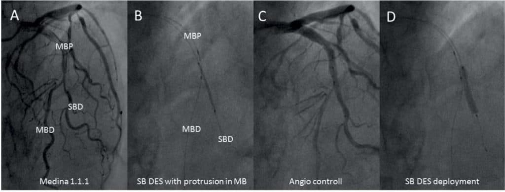
1.SB stent positioning with the adequate protrusion in MB – protrusion of the proximal stent segment into the MB (3-5 mm depending on bifurcati-on angle as performed in classical Crush technique)11 ensuring subsequent predictable crush and completebending of the protruding segment in only one direc-tion, opposite to the flow divider, thus leaving one single layer of stent struts at the ostium of the SB to be further crossed by the guidewire, and dilated by the SB balloon (Figure 2).
2.Proximal Side Optimization Technique (PSO) – if a two stent technique is applied, the bi-furcation involves by defi nition an important SB with extensive, long pathology. It should be considered that epicardial vessels present a tapering phenomenon, meaning discrepancy between distal as compared to proximal diameters. Since the SB stent size is based on the distal reference diameter, in long lesions there will be a definite size mismatch as compared to the ostial segment and stent deployment at nominal pressures can result in proximal under-expansion, leaving space for inadequate wire passage under the stent struts.
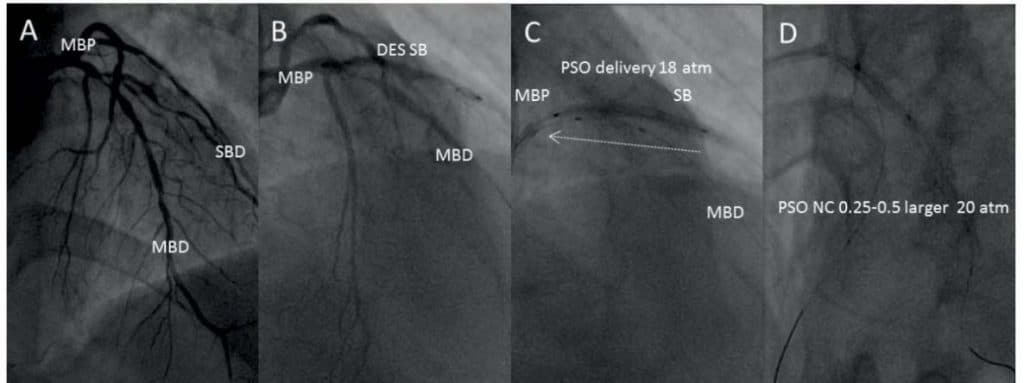
To avoid this complication we apply the PSO: After SB stent deployment at nominal pressure (Figure 3, A, B), the delivery balloon is pulled halfway back into the MB and second inflation at a higher pressure (~4-6 atm above nominal) is performed (Figure 3, C). Sub-sequently, a non-compliant (NC) balloon, 0.25-0.5 mm bigger than stent delivery is used to post dilate the implanted stent, after which is positioned half in MB and half in SB, and high-pressure infl ation is performed (Figure 3, D).
3.Crush – we suggest to crush the SB stent using the Proximal Optimisation Technique (POT) short balloon and using the POT technique14 – by infl ating an appropriately sized short NC balloon in the MV just proximal to the flow divider (Figure 4).
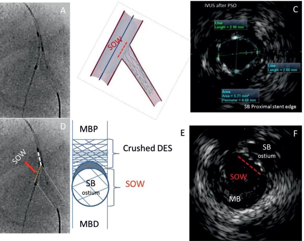
These three steps aim to: A – adequate protrusion – exclude the need to re-cross the crushed stent in the very proximal cell; any cell is perfect to be crossed at this time, B-effortless rewiring of the SB stent after crushing, C – PSO- proximal SB stent accommodation to the larger diameter with adequate strut apposition at the periostial segment, avoiding wire advancement and balloon delivery under the malapposed struts, D-POT-crushing the SB with a POT balloon in a POT manner creates a concave orientation of the bentstent in the SB ostium towards the SB, thus facilitating the wire easy ”slip” inside while recrossing.Briefl y, these three steps increase the space of op-timal wiring (SOW) of the SB stent after crushing (Figure 5)15. The following steps of the DK procedure are per-formed as described above, requiring less effort, time, and materials and bringing more reliable procedure and finally better acute and long term results.
DISCUSSION
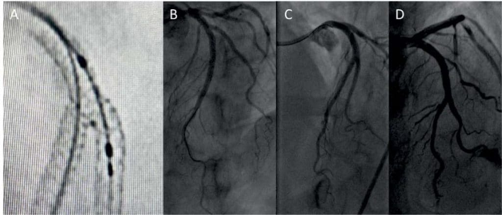
The promising results of SIRIUS and RAVEL trials16,17, reporting a substantial reduction in the rate of restenosis with the first generation of DES encouraged interventional cardiologists to approach progressively more complex lesions. Thus, meeting this demand, Dr. Colombo described the two stent crush technique to treat a bifurcation lesion11. The technical difficulty re-lied on the need to cross the 2 layers of thick stent struts (Cypher – 135 μm) for a final KBI. This tech-nique followed further refinement with the modification of Dr. Collins – it consisted of high-pressure NC balloon inflation at the ostium of the SB stent after Crush and before MB stent implantation18. This technique aimed to facilitate SB access after MB stenting. Bifurcation stenting gained further improvement by application of the Proximal Optimization Technique (POT), described by Daremont14, two-step KBI by Or miston19 etc. A further modification of the technique was the evolution of the mini-crush double KB – DK Crush described by Dr. Shao-Liang Chen12. Still, all the described optimizations were done after the SB stent Crush and the risk of first rewiring remains the same. The gaps in stent scaffolding occur usually on the side of the SB stent opposite to the crushed segment, as a consequence of balloon dilatation following the SB wire that exited MB stent and re-entered the SB stent after a course outside the SB stent struts. This phe-nomenon was observed by Ormiston, using microcomputed tomographic imaging of bench deployments and reported in 2008 (Figure 6)19. We repeated this simulation on a silicone phantom. We observed and reported, that by applying our modifications, the phenomenon of gaps in SB stent scaffolding was no more present20. Our modification is the only one that aims SB stent optimization before crush thus ensuring wide access to SB after crush due to increased SOW, which consequently reduces substantially the risk of inadequate wire passage. Therefore it was acknowledged by the European Bifurcation Club and mentioned in the cur-rent Consensus Document white paper21. The modification comes from an analysis of a large number of bifurcation stenting in our high volume center (>2300 PCI/year in 3 operators), and in our experience reduced considerably time, effort, and procedure costs in this complex subset of PCI (Figure 7).

Figure 1. Graphic representation of the risks of Side Branch (SB) stent crush in unpredictable fashion and subsequent guiding wire crossing under stent struts. A- SB stent deployed with minimal protrusion in main branch (MB) at nominal pressure (NP) with insufficient expansion and malapposition to the SB ostium due to vessel tapering phenomenon. B- SB stent crushed in unpredictable fashion. C- different possibilities of guiding wire crossing: green continuous line in the right way, red and purple dotted lines under the SB stent struts due to gaps in the ostial segment of the SB.

Figure 2. First step-SB stent positioning with adequate protrusion in MB. A- LAD-Diag MEDINA 1,1,1 bifurcation lesion treated with DK-Crush technique. B- SB DES positioned with sufficient protrusion in the MB (3-5 mm) and a NC balloon is positioned in the MB. C-Angio control of the SB stent positioning. D- SB stent deployment.

Figure 3. Second step- Proximal Side Optimization. A- LAD-Diag MEDINA 1,1,1 bifurcation lesion treated with DK-Crush technique. B- SB stent deployment. C- The delivery balloon is pulled halfway back into the MB and second inflation at a higher pressure (~ 4-6 atm above nominal) is performed. D- A non-compliant (NC) balloon, 0.25-0.5mm bigger than stent delivery is used to post dilate the implanted stent, after which is positioned half in MB and half in SB, and high-pressure inflation is performed.

Figure 4. Crush the SB stent using the Proximal Optimisation Technique (POT). A- “stent boost” imaging showing the distal marker of the short Non Compliant (NC) balloon in the MV just proximal to the fl ow divider (red arrow). MBP- Main Branch Proximal, MBD- Main Branch Distal, SBD- Side Branch Distal. B- Inflation of the short NC balloon at high pressure (20 atm). C- “stent boost” imaging after crushing of the SB stent, showing SB stent well expanded and is bent forming a monolayer in front of the ostium of SB in a concave shape.

Figure 5. The concept of the Space of Optimal Wiring (SOW). A, B, D- seen in “stent boost” showing a large space for wire crossing (yellow dotted line). C- IVUS image of the ostial segment of SB stent after PSO- confirming adequate expansion and apposition to the vessel wall and large cross-sectional area. E- Schematic representation of the SB stent after crushing “en face”- showing the stent strut monolayer and large crossectional SOW. F- IVUS image of MV after SB stent crush, showing large SOW at SB ostium.

Figure 6. Graphic representation of micro-computed tomographic imaging of bench stents deployments using Crush technique. Arrows showing the risk of gaps as a consequence of SB stent distortion due to crush in unpredictable fashion with subsequent wire and balloon passage under SB stent struts.

Figure 7. Final “stent boost” imanging and angiographic results. A- “stent boost” imanging and B, C, D-final angiographic results after applying the PSO modification showing perfect stent expansion, apposition, absence of gaps.
CONCLUSION
PSO is a promising technical iteration during crush stenting. Further imaging studies with phantom mi-crophotography and in-vivo intracoronary imaging are anticipated to further enlight on the beneficial effects on SB optimization. For now, single-center experience and opinion are that PSO allows the operator to optimize the SB ostium by 1) enabling sufficient strut apposition at the carina 2) eliminating the risk of SB DES distortion, 3) increasing the space of optimal re-wiring, 4) avoiding guidewire advancement in the peri-strut area. Although we describe this technical iteration during crush stenting, PSO can be widely applied also in other 2-stent techniques such as TAP and Cu-lotte. The technique is adding no additional equipment and saves time and effort to the operator and gives better results.
Abbreviations
DES, drug-eluting stent; SB, Side Branch; DK-Crush, double kissing Crush; MB, Main Branch; PSO, Proximal SB optimization; KBI, kissing balloon inflation; POT, Proximal Optimisation Technique; NC, Non Compliant; IVUS, intravascular ultrasound; SOW, Space of Optimal Wiring; TAP, T-stenting and small Protrusion.
Conflict of interest: none declared.
References
1. Chatzizisis Y.S., Jonas M., Coskun A.U., et al. Prediction of the local-ization of high-risk coronary atherosclerotic plaques on the basis of low endothelial shear stress: an intravascular ultrasound and histo-pathology natural history study. Circulation 2008;117:993–1002.
2. Gwon H.C., Choi S.H., Song Y.B., et al. Long-term clinical results and predictors of adverse outcomes after drug-eluting stent implantation for bifurcation lesions in a real-world practice: the COBIS (Coro-nary Bifurcation Stenting) registry. Circ J 2010;74:2322–2328.
3. Serruys PW. The treatment of coronary bifurcations: a true art form. EuroIntervention 2015;11 Suppl V:V7.
4. Ford TJ, McCartney P, Corcoran D, et al. Single-Versus 2-Stent Strategies for Coronary Bifurcation Lesions: A Systematic Review and Meta-Analysis of Randomized Trials With Long-Term Follow-up. J Am Heart Assoc. 2018 May 25;7(11):e008730.
5. Di Gioia G, Sonck J, Ferenc M, et al. Clinical outcomes following coronary bifurcation PCI techniques: a systematic review and net-work meta-analysis comprising 5,711 patients. J Am Coll Cardiol Intv. 2020 Jun, 13(12):1432-1444.
6. Neumann FJ, Sousa-Uva M, Ahlsson A, et al. 2018 ESC/EACTS Guidelines on myocardial revascularization; ESC Scientific Docu-ment Group. Eur Heart J. 2019 Jan 7;40(2):87-165. doi: 10.1093/ eurheartj/ehy394.
7. Young Bin Song, Joo-Yong Hahn, Jin-Ho Choi, Seung-Hyuk Choi, Sang Hoon Lee, Hyeon-Cheol Gwon. Bifurcation Angle And T-Stenting And Small Protrusion (TAP) Bifurcation Percutaneous Cor-onary Intervention Technique J Am Coll Cardiol. 2010 Mar, 55 (10 Supplement) A199.E1875. doi: 10.1016/S0735-1097(10)61876-1.
8. Kim HY, Doh JH, Lim HS, et al.: Identifi cation of Coronary Artery Side Branch Supplying Myocardial Mass That May Benefit From Re-vascularization. JACC Cardiovasc Interv. 2017 Mar 27;10(6):571-581.
9. Lassen JF, Holm NR, Banning A, Burzotta F, Lefèvre T, Chieffo A, et al. Percutaneous coronary intervention for coronary bifurcation dis-ease: 11th consensus document from the European Bifurcation Club. EuroIntervention 2016;12:38–46.
10. Chen SL, Xu B, Han YL, et al. Comparison of double kissing crush versus Culotte stenting for unprotected distal left main bifurcation lesions: results from a multicenter, randomized, prospective DK-CRUSH-III study. J Am Coll Cardiol 2013;61:1482–1488
11. Colombo A, Stankovic G, Orlic D, Corvaja N, Liistro F, Airoldi F, et al. Modified T-stenting Technique With Crushing for Bifurcation Le-sions: Immediate Results and 30-day Outcome Catheter Cardiovasc Interv 2003 Oct;60(2):145-51. doi: 10.1002/ccd.10622.
12. Zhang JJ, Chen SL. Classic crush and DK crush stenting techniques. EuroIntervention 2015;11 Suppl V:V102-5. doi: 10.4244/EIJV11S-VA23
13. Chen X, Li X, Zhang JJ, Han Y, Kan J, Chen L, Qiu C, Santoso T, Paiboon C, Kwan TW, Sheiban I, Leon MB, Stone GW, Chen SL; DKCRUSH-V Investigators. 3-Year Outcomes of the DKCRUSH-V Trial Comparing DK Crush With Provisional Stenting for Left Main Bifurcation Lesions. JACC Cardiovasc Interv. 2019 Oct 14;12(19):1927-1937. doi: 10.1016/j.jcin.2019.04.056.
14. Darremont O, Leymarie JL, Lefè vre T, Albiero R, Mortier P, Louvard Y. Technical aspects of the provisional side branch stenting strategy. EuroIntervention. 2015;11(suppl V):V86–V90. doi: 10.4244/ EIJV11S-VA19
15. Lavarra F. Proximal Side Optimization: A modifi cation of the dou-ble kissing crush technique. US Cardiology Review 2020;14:e02. doi. org/10.15420/usc.2020.07
16. Moussa I, Leon MB, Baim DS, O’Neill WW, Popma JJ, Buchbinder M, Midwall J, Simonton CA, Keim E, Wang P, Kuntz RE, Moses JW. Impact of sirolimus-eluting stents on outcome in diabetic patients: a SIRIUS (SIRolImUS-coated Bx Velocity balloon-expandable stent in the treatment of patients with de novo coronary artery lesions) substudy. Circulation 2004 May 18;109(19):2273-8. doi: 10.1161/01. CIR.0000129767.45513.71
17. Abizaid A, Costa MA, Blanchard D, Albertal M, Eltchaninoff H, Gua-gliumi G, Geert-Jan L, Abizaid AS, Sousa AG, Wuelfert E, Wietze L, Sousa JE, Serruys PW, Morice MC; Ravel Investigators. Sirolimus-eluting stents inhibit neointimal hyperplasia in diabetic patients. In-sights from the RAVEL Trial. Eur Heart J. 2004 Jan;25(2):107-12. doi: 10.1016/j.ehj.2003.11.002.
18. Collins N, Dzavik V. A modified balloon crush approach improves side branch access and side branch stent apposition during crush stenting of coronary bifurcation lesions. Catheter Cardiovasc Interv. 2006 Sep;68(3):365-71. doi: 10.1002/ccd.20791.
19. John A Ormiston, Mark W I Webster, Bruce Webber, James T Stewart, Peter N Ruygrok, Robert I Hatrick .The “crush” technique for coronary artery bifurcation stenting: insights from micro-computed tomographic imaging of bench deployments JACC Cardiovasc Interv. 2008 Aug;1(4):351-7. doi: 10.1016/j.jcin.2008.06.003.
20. Franceso Lavarra. Proximal Side-Branch Optimization in Crush stenting. A step-by-step Technical Approach in a Silicone Phantom Model. Cardiovascular Revascularization Medicine. 2020; 10.1016/j. carrev.2020.07.037.
21. Francesco Burzotta, Jens Flensted Lassen, Yves Louvard, et al. Eu-ropean Bifurcation Club white paper on stenting techniques for pa-tients with bifurcated coronary artery lesions. Catheter Cardiovasc Interv. 2020 Jun 24. doi: 10.1002/ccd.29071. Online ahead of print. PMID: 32579300 DOI: 10.1002/ccd.29071.
 This work is licensed under a