Mirela-Anca Stoia1,2, Anca Daniela Farcas1,2, Calin Homorodean1,3, Dan Mircea Olinic1,3
1 First Medical Clinic, „Iuliu Hatieganu” University of Medicine and Pharmacy, Cluj-Napoca, Romania
2 Department of Cardiology, Emergency County Clinical Hospital, Cluj- Napoca, Romania
3 Department of Cardiology No.2, Interventional Cardiology, Emergency County Clinical Hospital, Cluj-Napoca, Romania
Abstract: Background – Carotid artery stenosis (CAS) is related to the extent of coronary artery disease (CAD) and multi-vessel CAD is an independent predictor for CAS. Simultaneous ATS lesions of the carotid arteries (carotid intima-me- dia thickness-CIMT) increases and extracranial cervical stenosis due to carotid plaques) and coronary circulation indicates the extent of ATS burden. Aim – study of comparative cardiovascular risk factors (CVRF) profile, CIMT, CAS, and CAD patterns in acute coronary syndrome (ACS) and in stable CAD patients. Method – laboratory tests for CVRF, arterial cervical US parameters measurements, and coronary angiography evaluation. Results – ACS patients had more diabetes mellitus (72.2% DM) and hypertriglyceridemia (56.4% HTgl), while stable CAD patients were more smokers (53.5%) and hypertensive (78.7% HT). ACS patients had increases CIMT (1.07±0.26 mm vs. 0.7±0.2 mm). Stable CAD patients had more CAS (63.7% vs. 33.3%), but in ACS patients, the severity of CAS increases with the number of coronary artery lesions. Con- clusions – CAD patients have particular CVRF profile, different for ACS patients and stable CAD patients and these could be related to their specific CAS-CAD patterns. In ACS patients, increased CIMT, and carotid stenosis are associated with the presence, the extent, and the severity of coronary stenosis, and might be useful in estimating ATS burden in patients with multi-vessel disease, in particular with left anterior descending coronary artery lesion.
Keywords: coronary artery disease, carotid artery stenosis, carotid intima media thickness, cardiovascular risk factors.
INTRODUCTION
Atherosclerosis (ATS) is a systemic, extensive, diffuse, and progressive inflammatory, metabolic, and immune vascular disorder, involving multiple arterial beds. Pro- gressive carotid lesions are reportedly a stronger pre- dictor of imminent myocardial infarction (MI) than of future stroke events1,2. Carotid stenosis is related to the extent of CAD and multi-vessel coronary lesions had shown to be an independent predictor for CAS3. The presence of symptoms of carotid disease is alar- ming and certainly correlates with poor outcome, but the absence of symptoms is not a particular security for a good outcome4.
ATS changes in carotid morphology include CIMT increases, correlated with endothelial dysfunction, carotid plaque formation and deposition leading to arterial stenosis, also present simultaneous in the co- ronary arteries territory. Concomitant ATS lesions of the extracranial carotid and coronary circulation indicates the extent of ATS burden and portend an adverse prognosis in various clinical settings, including asymptomatic individuals, stroke patients, and patients undergoing coronary artery bypass surgery (CABG)1. There are limited studies focused on the prevalence of asymptomatic carotid disease in association with CAD other than candidates for major cardiac surgery, indi- cating a strong link with multi-vessel CAD5. We did not found specific observations from studies regarding particularities of the relationship between coronary and cervical arteries lesions in patients admitted with ACS and comparative differences with stable CAD patients.
SUBJECTS AND METHODS
The study was performed in the Cardiology De- partment of Emergency County Clinical Hospital of Cluj-Napoca. After signing an informed consent, we enrolled 167 consecutive patients (pts.) without any anamnestic clinical carotid artery symptomatology, and without any documented history of established or transient stroke: 87 with ACS [10 p. with ST elevati- on (STEMI) and 77 p. without ST elevation (NSTEMI)] and 80 with stable CAD. The NSTEMI criteria accor- ding to guidelines6 definition were: acute chest pain [prolonged (>20 min) angina pain at rest, or new onset (de novo) angina, or recent destabilization of previo- usly stable angina (crescendo angina)], and elevation of cardiac troponin cTnI above the 99th percentile of the laboratory cut-off value (0.02 ng/l). The stable CAD patients selected had inducible ischemia during exercise stress tests and no elevation of cTnI. In all patients we performed clinical evaluation, 12 lead electrocardi- ogram (ECG), hemodynamic monitoring, and labora- tory tests for CVRF (glucose, cholesterol, triglyceride serum levels). CVRF were defined according to the criteria in the current guidelines. Smoker status re- ferred to the active smoker. The presence of dysli- pidaemia was defined by increased >200 mg of total cholesterol and/or increased >150 mg of triglycerides serum levels7. Diabetes diagnosis was made prior to inclusion in the study.
All patients underwent imaging vascular duplex ul- trasound (US) evaluated cervical arterial lesions, topo- graphy and hemodynamic significance of CAS. The US scan was made on Hewlett Packard HP Sonos 4500 machine using a 7.5 MHz linear transducer, by a single specialist doctor examiner (certified for cardiovascu- lar US competence) who was blinded to clinical and coronary angiographic data, using a described below protocol The patients with ACS followed coronary angiography evaluation and the cervical arteries US evaluation was made in the first 24-72 hours from ad- mittance.
Arterial cervical US parameters were measured: CIMT measurements were made in the bilateral com- mon carotid artery (CCA), in longitudinal projecti- on (the lumen intima and media-adventitia interfaces were clearly defined), between two parallel echogenic lines corresponding to the distance between the le- ading edge of the luminal echo (first bright line) and the leading edge of the media-adventitia echo (second bright line), on the posterior-medial wall, standard 2 cm before the bulb carotid bifurcation, in a free of plaque region, using manual reading system. CIMT was calculated as the average of the CIMT for left and right CCA. The average values measured were compared with normal values considered <0.75 mm8. The CCA- IMT value >1.2 mm was considered as ATS plaque6. The estimation of hemodynamic significance of CAS or the degree of luminal narrowing was assessed using the following elements: morphology criteria provided by duplex color cross-section carotid luminal planime- try in B-mode US with the calculation of the stenosis area (%) by the formula: total vessel area-duplex color permeable area/total vessel area, and hemodynamic criteria: peak-systolic velocity (PSV), end-diastolic ve- locity (EDV), intra-stenosis/pre-stenosis PSV ratio9-12. Stenosis between 50-70% was considered moderate CAS and was defined by the follow arterial US para- meters: PSV=1.25-2.3 m/s, EDV <0.4 m/s, PSV intra- stenosis/pre=2-3, and by corresponding percentage reduction of the duplex color area reported to the functional duplex values9-12. Stenosis >70% was de- fined by the follow values: PSV >2.3 m/s, EDV >0.4 m/s, PSV intra-stenosis/pre-stenosis ratio >3 and by corresponding percentage reduction of the functional area reported to the functional duplex values9-12. Ar- terial luminal occlusion was defined by the presence of a total occluding mass with the absence of color and pulsate duplex flow. Stenosis >70% and occlusion were considered severe CAS9-12. Variation coefficients of the measurements for the examiner were <5%.
Coronary angiography was performed in emergen- cy for STEMI patients and in an emergency elective manner for NSTEMI patients in the first 24-72 hours after admittance. The stable CAD patients were selec- ted having positive inducible myocardial ischemia on treadmill exercise stress electrocardiogram test and they underwent coronary angiography. By coronary angiography, we evaluated the topography and the hemodynamic significance of coronary artery lesions. Coronary angiography was performed by standard te- chniques on Siemens angiography system 2000. The severity of coronary lesions was determined by visual estimation at the discretion of the interventional car- diologist performing the procedure. Clinically signifi- cant CAD was defined as the presence of a coronary lesion resulting in a lumen diameter stenosis >70% in a major epicardial artery (left anterior descending ar- tery (LAD), left circumflex artery (Cx), right coronary artery (RC) or one of its major branches). Clinically significant left main disease (LMD) was defined as a lumen diameter stenosis >50%. Patients were strati- fied according to the number of involved vessels as follows: normal coronaries or non-obstructive CAD (individuals not meeting the criteria for clinically sig- nificant CAD), and 1VD, 2VD, 3VD/LMD (significant lesions in 1, 2, and 3 vessels or left main disease, re- spectively). The study methodology was approved by the Ethical Committee. Data was analyzed using SPSS version 2.0. Categorical variables were expressed as proportions and percentages and continuous variables were expressed as means ± standard deviation. Stu- dent T-test was used to compare PSV, EDV, and intra/ prestenosis PSV ratio. For categorical variable Chi- square test was used. p<0.01 was consider statistically significant.
RESULTS
Descriptive characteristics established for the two groups and for all subjects and the CVRF profiles are presented in Table 1. The distribution of coronary ar- teries lesions diagnosed by angiography examination in ACS patients is represented in Figure 1.
In ACS patients, the median value of CIMT was sig- nificantly higher than in stable CAD patients, in which CIMT values were around normal range. CIMT was significantly higher in 3VD patients than in 2VD of ACS patients. CIMT was similarly increased in1VD patients and 2VD patients, as well as in patients without signifi- cant coronary stenosis. Some of the ACS patients had a “normal” CIMT value (i.e. Fig. 2).
The prevalence of overall CAS >50% was higher in stable CAD pts. than in ACS pts. Both prevalence of CAS=50-70% and prevalence of CAS >70% or carotid occlusion were higher stable CAD pts. in comparison with ACS pts. (i.e. Table.2). The prevalence of multi- vessel coronary stenosis (2VD and 3VD) was higher in stable CAD patients comparative with ACS patients, while the prevalence of LAD coronary artery lesions was higher in ACS patients (75.8%) in comparison with stable CAD. CAS >50% was significant higher in stable CAD patients comparative with ACS patients. (i.e. Table 2).
The prevalence of overall CAS >50% in 3VD, 2VD and 1VD of ACS patients is represented in Table 3 and Figure 3.
The prevalence of moderate and severe CAS in 3VD, 2VD and 1VD of ACS patients are presented in Figure 4. The prevalence of CAS >50% was significant higher in 1VD and 2VD of ACS patients with a LAD coronary involvement, than in those with Cx and/or RC lesions (i.e Fig.5) In unadjusted linear regression analysis cholesterol, HT, smoking status and age were positively associated with the lesions in stable CAD patients. Cholesterol, triglycerides serum levels, the presence of diabetes, and smoking status were positively associated with the carotid lesions in ACS patients (i.e. Table 4).
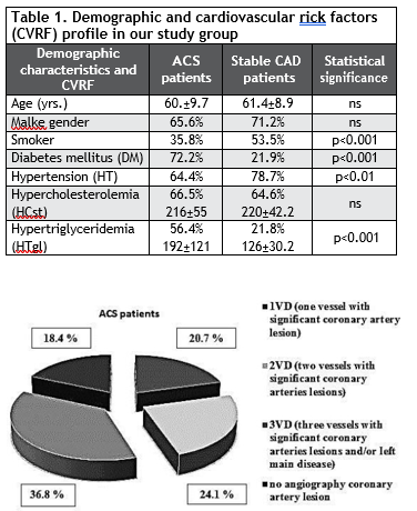
Figure 1. Coronary artery lesions repartition in ACS patients.
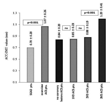
Figure 2. Comparative IMT values measured on CCA in study group.
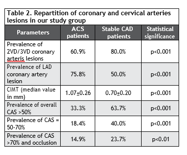
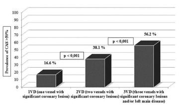
Figure 3. Comparative repartition of overall CAS >50% related to the dis- position of coronary arteries lesions in ACS patients from our study group.
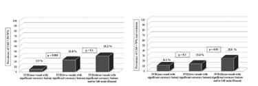
Figure 4. Relationship between the prevalence of moderate, respectively severe CAS, and coronary arteries lesions significances in ACS patients from our study group.

Figure 5. The prevalence of multisite CAS related to the multi-vessel cor- onary arteries lesions significances in ACS patients and the higher preva- lence of CAS >50% in 1VD and 2VD of ACS patients with LAD lesions from our study group.


DISCUSSIONS
The data from studies shows that the patients with coronary-carotid tandem lesions are at middle age (45-64 yrs.) and with a slightly prevalence of female gender, especially for the patients with critical CAS13. Patients from our study were majority male, and not very elderly, with similar values of median age years. ACS patients had more DM, and hypertriglyceridemia, while stable CAD patients were more smokers and had more HT. The prevalence of hypercholesterole- mia was almost equally high. The data regarding the prevalence of multiple CVRF are very different in vari- ous study, explain by the diversity of patients groups. In REACH registry the proportion of multiple CVRF was 18.3%, less than in our group14,15. There are stu- dies emphasized that none of these CVRF (HT, DM, dyslipidemia, obesity, smoking, family history) can be a potential predictor of CAS. Other studies showed that peripheral vascular disease (PAD), previous ische- mic stroke, smoking, and female gender, are markers associated with important CAS16,17.
The prevalence of smoking and HT did exceed 80% in CAD population from some studies13,17. In others, variables such as HT, or hyperlipidemia were not sta- tistically different3. The REACH registry revealed also high prevalence of HT (80.3%), hypercholesterolemia (77%), DM (38.3%), but not so high prevalence of obe- sity/ hypertriglyceridemia (29.9%) and smoking (13%) in CAD patients14,15. These differences could result from the ethnic variety of REACH patients enrolled from many countries, from the fact that CAD patients were not separated in ACS and stable CAD, implying particular different CVRF patterns, and from some specific CVRF exposure in Romanian citizen.
The smoking, HT, hypercholesterolemia and DM are associated with ATS of extracranial carotid arte- ries. Diabetic patients with CAD often have a combination of elevated triglycerides and low level of HDL than high-level of LDL cholesterol3,13,17,18. We might make the assumption that the higher prevalence of DM in our ACS patients might be prone to a more pro-inflammatory, and pro-thrombotic status associa- ted with the worsening of endothelial dysfunction and increases CIMT, while smoking and HT seems to be more related to a progressive fibro-calcific ATS evo- lution. These hypotheses do not suggest a one to one correspondence between carotid and coronary artery lesions formation and development. The correlated presence of tandem carotid and coronary artery le- sions do not necessarily share the same associations with similar CVRF. Differences in ATS lesions preva- lence and incidence in both arterial systems may be related also to marked morphologic, hemodynamic and geometric differences between coronary and ca- rotid arteries19.
Some studies have shown that CIMT could be a re- liable marker for CAD and might improve the predic- tion ability for CV events risk over and above conven- tional CVRF20. The use of CIMT for assessment of CV event risk has a number of limitations too. Although increased CIMT has been shown to be a strong pre- dictor of incident MI in overall population >55 years, it is not a good predictor for the extent and the severity of CAD. The normal CIMT cannot predict the absen- ce of significant CAD. There are several explanations like: considerable heterogeneity among various stu- dies regarding the characteristics of the participants, CIMT methodology and measurement protocols, and the interpretation of the results21-23.
The uppers values of normal CIMT varies with the site of carotid measurement, increasing from ICA to ACC, but not significant (0.62 to 0.74 mm)21-23. The majority of CIMT studies agree that a CIMT value >0.75 mm could be accepted as pathological increa- ses. On other hand a CIMT value >1.2 mm (1.1-1.5 mm in different studies) can be considered as an ATS deposition (carotid plaque)22. In European countries, it is recommended that measurement of CIMT sho- uld be performed in a region free of plaque and the distinction between CIMT and carotid plaque should be clearly made. CIMT normal value increases with progression of age, but this interval values between 0.7-1.2 mm are mostly accepted as normal regardless of age21-24.
We did not found significant studies regarding the particularities of CIMT values significances and CAS profiles in ACS patients comparative with stable CAD patients. CIMT values significant increases in CAD pa- tients with multi-vessel lesions (≥2VD)25. In this con- text, we can stipulate the fact that increases values of CIMT in ACS patients (with an slowly ascending trend from 1VD to 2VD and respectively with a sig- nificant increasing bound to 3VD) might reflects a more” aggressive” coronary and carotid arteries ATS evolution, possible related to the high presence of DM, inflammation and to the endothelial dysfunction linked to arterial stiffness, which, by itself, is associa- ted with adverse CV outcomes. In ACS patients, the- re is a stepwise increase in carotid lesions with the increasing severity of CAD, and carotid lesions may reflect a diffuse CAD. The extent of non-obstructive CAS lesions has been found to be a strong predictor of coronary events, independent of severity and the number of flow-limiting coronary lesions21. In support of what has been already stipulated, even our ACS patients with no significant coronary lesions had CIMT values higher than normal. In opposition, stable CAD patients seems to have a more slowly and progressive coronary and carotid ATS evolution, which “allows” the development of an extensive and multisite vessels ATS lesions possible related to their particular meta- bolic risk profile (smoking, HT, HCst).
The natural history, risk factors patterns, and cardi- ac and cerebral events prediction are different for “ca- rotid plaque”, which is a focal structure en-croaching into the arterial lumen, and the “traditional” IMT, which is measured in a region free of plaque, where- as both share some common ATS risk factors20. The presence of carotid lesion is associated with incident positive and more rapid and diffuse calcification pro- gression of coronary artery lesions and, from a sta- tistical point of view, more significant than DM and smoking18. Like CIMT, carotid plaque reflects the ove- rall ATS burden, and, as compared with IMT, better predicts CV death and nonfatal MI22. The assumptions aforesaid could explain also the particularities of CAS in our group study. The stable CAD patients had a higher prevalence of CAS >50% comparative with ACS patients, probably due to their longer ATS evolution. Further stable CAD patients had significant more mo- derate CAS than ACS patients, but regarding severe CAS, there was only a moderate prevalence in favor of stable CAD patients. In stable CAD patients group we found more moderate than severe CAS, while in ACS patients was a relatively similar proportion of mode- rate and severe CAS.
The prevalence of asymptomatic CAS in CAD pati- ents is variable in different studies: 10% in REACH re- gistry (started in 2003)13,14, 7-18% in ESC PAD guide- lines (2017)24, in others 17-22%13, but this proportion can be greater>30% in high-risk CAD patients prone to ACS. But in discrepancy, the prevalence of CAD in CAS patients is higher: 39-61% in ESC PAD guide- lines (2017)24, 28-40% in other studies26 so the relati- onship is reciprocal unequal and need further studies. However the overall rates for CAS in CAD patients varies from 5% to 30. In REACH registry, at 4-year follow-up, the presence of carotid ATS was associated with a 22% increase in the risk of coronary events; consistent finding across all patient subsets, including patients with or without pre-existing CAD27. The pre- sence of carotid plaque at baseline is associated with a 7-fold increase in the risk of cardiac death and a 2-fold increase in the rates of the composite end point (all- cause mortality, MI, and stroke)28.
The presence of carotid ATS is directly related to the extent of CAD, though the prevalence of mode- rate and severe CAS in patients with CAD is varia- ble, but in general lower than our findings1. There are several explanations: first that in many studies the percentage of CAS is calculated by B-mode US lon- gitudinal diameter narrowing, not by transversal pla- nimetry area reduction (which is more accurate)8-11. Second, from our previous observations, we found that carotid PSV between 1.25-1.7 m/s can indicate sometime no significant stenosis (<50%), that why is better to add carotid EDV and PSV intra/pre-stenosis ratio16,29-33. Third, there is a discrimination between carotid ATS (non-stenotic carotid plaque <50%) and CAS (stenotic carotid plaque >50%). The prevalen- ce of CAS>50% in our SCAD patients was 63.7%, in other studies has varied from 5,8% to 80%, depending if carotid ATS or CAS (defined both as well as carotid disease) was taken into consideration15,20. Moreover there are some differences regarding the calculation of CAS hemodynamic significance in USA (NASCET, PSV intra/post-stenosis ratio34) and in Europe (ECST, PSV intra/pre-stenosis ratio35) and depends which one was used in the studies. Four, the definition of “significant” CAS was different across the studies, in many studies were reported the overall carotid plaque, irrespective to the stenosis degree, while in others moderate CAS (50-70%) or only CAS >70% were considered. In fifth place, beside the difference in methodologies, there was inhomogeneity in studies group, sample size and in the various ethnic compositions. There were very different categories of studies inclusion criteria like: asymptomatic individuals, patients undergoing CABG with or without prior MI or stoke, so with stable CAD, but rarely ACS patients36.
The proportion of CAS increases with the number of coronary lesions: 5-14% (from studies) in CAD pa- tients with 1VD (16.6% CAS >50% in our study), 13- 21% in 2VD (38.1% in our study), 17-36% in 3 VD, and 31-40% in LMD (56.2% in our study including 3VD and LMD)1,37-39, although there are studies who found that CAS is correlated with LMD, and it is not correlated with multi-vessel CAD15. There are studies showing that LMD patients had more CAS than 3VD pati- ents1,15,25, in others the proportion of CAS was similar in LMD and 3VD CAD patients36. Multiple CAS was found in 23.8% of 2VD and 50% of 3VD ACS patients, so often multisite CAD means multiple CAS. But we have to mention that our study percentages are rela- ted to ACS patients and they are 1 fold greater than the reported numbers, probably related to the same imbalanced CVRF profile and to the modality of ATS evolution in ACS patients. Another thing is that the greater percentage of CAS arrives from older studies (around 2000)37, while the smaller percentage is from more recent studies (2005-2010)1,38,39. The explanati- on might be better and intensive prevention medical therapy of CVRF1.
CAS was more severe in 1VD and 2VD ACS pa- tients, while 3VD ACS patients had more moderate CAS. Moreover 1/2 of the 3VD and 1/3 of the 2VD ACS patients had multiple CAS, aspects that might be related to cyclic (inflammation-calcification) progres- sive evolution of ATS. Not least, we found another interesting correlation between the presence of pre- dominant LAD lesions and the prevalence of CAS in 1VD and 2VD ACS patients: ACS patients with LAD lesions had 2 fold higher CAS than ACS patients with Cx and/or RC lesions. We did not found this obser- vation in other studies and we just can stipulate that LAD lesion-CAS correlation could be related proba- bly to the pattern of ATS evolution in ACS patients and maybe to some hemodynamic particularities of LAD coronary circulation.
It is unclear why ATS lesions progress silently for extended periods of time and then suddenly undergo changes leading to plaque instability, rupture, throm- bosis, and acute clinical syndromes. These are not limited to a single “culprit” site, they are part of a multi-centric inflammatory process with many triggers and involving simultaneous multiple additional plaqu- es instability at distantly locations in the entire arte- rial system21,40. Inflammation may be the link between coronary and carotid plaque destabilization, reflected in elevated high sensitivity serum C-reactive protein (hsCRP), interleukin-6, adhesion molecule-121. The acute phase reactants reflect the systemic inflammati- on, but they are also active participants to the process of ATS plaques formation, and may be modulated by focal changes in the carotid arteries21. Maybe other new biomarkers related to coronary ischemia and in- creased incidence of CV events (neutrophil /lympho- cyte ratio, plasma leptin, resistin, TNF-a, adiponectin) or to cardiovascular stiffness in HT patients (serum ST-2 soluble) could be more specific for carotid and coronary ATS inflammation and might be added to the clinic, metabolic, and imagistic evaluation of ATS in order to increase prognostic value in polyvascular patients41-45.
LIMITATION OF THE STUDY
A limitation is the small number of subjects. Despite the fact that noninvasive assessment of CIMT and ca- rotid plaque burden is a marker for future coronary events, and although current guidelines on CVD pre- vention consider the presence of any degree of CAS a high-risk marker of cardiovascular events, routine carotid US Doppler screening is not currently endor- sed. Our data, however, did not propose to resolve the existing controversy regarding the reciprocal re- lationship between CAD and CAS, but some particu- lar aspects of CVRF profile and US carotid elements, in ACS patients in comparison with stable CAD have been highlights in our study. The prognostic relevan- ce of US characteristics of carotid plaques in patients with CAD, possibly in association with inflammatory markers, needs to be further clarified.
CONCLUSIONS
In our study, we found that CAD patients have parti- cular CVRF profiles, different for ACS patients (more DM and hypertriglyceridemia), and for stable CAD patients (more smoking and HT) and these specific fin- dings could be related to their specific CAS-CAD pat- terns. Stable CAD patients had more moderate than severe CAS, while in ACS patients was a relatively si- milar proportion of moderate and severe CAS. This is one of the first studies focused on CAS particularities in ACS patients. Increased CIMT, and carotid stenosis were associated with the presence, the extent, and the severity of epicardial coronary stenosis in ACS patients, and might be useful in estimating ATS bur- den in those patients with multi-vessel disease. Our study shows a direct correlation between increasing CIMT values and severity of coronary lesions in ACS patients, as a surrogate marker of acute CV events, and also for revascularization. We find that the high prevalence of CAS increases in parallel with the num- ber of the involved coronary arteries in ACS patients and had a specific correlation with LAD lesions. Inde- pendent predictors of severe CAS were: the presence of LMD or 3VD, increasing age, smoking status, DM. Although CAS is known as a risk factor for future ad- verse CV events in CAD patients, it might be a marker for diffuse ATS disease burden rather than a direct etiologic factor.
Conflict of interest: none declared.
References
1. Steinvil A, Sadeh B, et al. Prevalence and Predictors of Concomitant Carotid and Coronary Artery Atherosclerotic Disease. JACC Vol. 57, No. 7, 2011:779-83
2. Steinvil A, Sadeh B, et al. Impact of Carotid Atherosclerosis on the Risk of Adverse Cardiac Events in Patients With and Without Coro- nary Disease. Stroke, 2014;45:2311-2317
3. Avci A, Fidan S, et al. Association between the Gensini Score and Carotid Artery Stenosis. Korean Circ J, 2016;46( 5): 639 -645
4. Vranic H, Hadzimehmedagic A, et al. Critical Carotid Artery Steno- sis in Coronary and Non-Coronary Patients-Frequency of Risk Fac- tors. Med Arch. 2017; 71(2): 110-114
5. Fassiadis N, Adams K, et al. Occult carotid artery disease in patients who have undergone coronary angioplasty. Interactive CardioVascu- lar and Thoracic Surgery 7 (2008) 855–857
6. Baumgartner H, Gaemperli O, et al. 2015 ESC Guidelines for the management of acute coronary syndromes in patients presenting without persistent ST-segment elevation. European Heart Journal (2016) 37, 267–315
7. Piepoli MF, Hoes AW, et al. 2016 European Guidelines on cardiovas- cular disease prevention in clinical practice. European Heart Journal (2016) 37, 2315–2381
8. Peters SAE, den Rijter HM, Bots MI. Ultrasound protocols to mea- sure carotid intima-media thickness: one size does not fit all. J Am Soc Echocardiography, 2012;25;10:1135-37
9. Stoia M. Ecografia arterelor periferice şi a sistemului arterial carot- ido-vertebral în: Vida–Simiti L, et al. Explorări nonivazive în bolile cardiovasculare. Indrumător pentru studenţi şi rezidenţi. Editura Medicală Universitară „Iuliu Haţieganu” Cluj Napoca, 2011:196-218
10. Strandness E.D. Extracranial artery disease in Duplex scanning in Vascular Disease, Lippincott, Williamson and Wilkins, a Woter Klu- ver Company, Philadelphia, 2012;84-115
11. Dauzat M. L’examen ultrasonographique des axes carotidiens in Practique de l’ultrasonographie vasculaire par Dauzat M., Ed. Mas- son, Paris, 2002;96-177
12. Bekelis K, Labropoulos N, et al. B-mode estimate of carotid steno- sis: planimetric measurements complement the velocity estimate of internal carotid stenosis. Int Angiol, 2013;32(5):506-11
13. Vranic H, Hadzimehmedagic A, et al. Critical Carotid Artery Steno- sis in Coronary and Non-Coronary Patients-Frequency of Risk Fac- tors. Med Arch. 2017; 71(2): 110-114
14. Valentijn TM, Stolker RJ. Lessons from REACH Registry in Europe. Current Vascular Pharmacology, 2012;10:725-727
15. Suarez C, Zeymer U, et al. Influence of polyvascular disease on car- diovascular event rates. Insights from REACH Registry. Vascular Medicine, 2015;15(4): 259-65
16. Brenovic-Kircanski O, Panic D, et al. Role of risk factors in predic- tion of asymptomatic carotid artery stenosis in patients with coro- nary artery disease. Acta Medica Mediterranea, 2016, 32: 63
17. Stoia M.A., Farcas AD, et al. Comparative Analysis of Cardiovascu- lar Risk Profile, Cardiac and Cervical Arterial Ultrasound in Patients with Chronic Coronary and Peripheral Arterial Ischemia. Interna- tional Conference on Advancements of Medicine and Health Care through Technology, IFMBE Proceedings, vol 59. Springer, 2017 DOI: 10.1007/978-3-319-52875-513:53-56
18. Tolstrup JS, Hvidtfeldt UA, et al. Smoking and Risk of Coronary Heart Disease in Younger, Middle-Aged, and Older Adults.American Journal of Public Health. 2012; e1-e7
19. Polak FJ, Tracy R et al. Carotid artery plaque and progression of coronary artery calcium: the Multi-Ethnic Study of Atherosclerosis. J Am Soc Echocardiogr. 2013;26(5):548–555
20. Farcas AD, Vonica CL, et al. Non-alcoholic fatty liver disease, bulb carotid intima-media thickness and obesity phenotypes: results of a prospective observational study. Med Ultrason 2017:0, 1-7
21. Rie Y, Katakami N, et al. The Utility of Carotid Ultrasonography in Identifying Severe Coronary Artery Disease in Asymptomatic Type 2 Diabetic Patients Without History of Coronary Artery Disease. Diabetes Care, 2013; 36:1327–1334
22. Kasliwal RR, Kaushik M, et al. Carotid Ultrasound for Cardiovascular Risk Prediction: From Intima media Thickness to Carotid Plaques. Indian Acad Echocardiogr Cardiovasc Imaging 2017;1:39-46
23. Hariawan H, Harris M, et al. Correlation between Carotid Intimal- Media Thickness and Coronary Artery Disease Severity in Stable Coronary Artery Disease Patients. Acta Cardiologia Indonesiana, 2018, 3(2):81-88
24. Aboyans V, Ricco JB, et al. 2017 ESC Guidelines on the Diagnosis and Treatment of Peripheral Arterial Diseases, in collaboration with the European Society for Vascular Surgery (ESVS). European Heart Journal (2017) 00,1–6
25. Murray F. Matangi MD. Carotid Artery Thickness and Plaque Quan- tified by Carotid Ultrasound is Associated with Angiographic Coro- nary Stenosis. Abstract P2-46, 23nd Annual Scientific Sessions of the ASE, 2012
26. Dos Reis PFF, Linhares PV, et al. Approach to concurrent coronary and carotid artery disease: Epidemiology, screening and treatment. Rev Assoc Med Bras 2017; 63(11):1012
27. Sirimarco G, Amarenco P, et al; REACH Registry Investigators. Ca- rotid atherosclerosis and risk of subsequent coronary event in out- patients with atherothrombosis. Stroke. 2013;44:373–379
28. Park HW, Kim WH, et al. Carotid plaque is associated with in- creased cardiac mortality in patients with coronary artery disease. Int J Cardiol. 2013;166:658–663
29. Stoia M. Ultrasonografia vasculară-capcane sau nu? în Gherman CD. Angiologia şi chirurgia vasculară. Actualităţi şi perspective. Editura Cărţii de Ştiinţă, 2016, p.67-80
30. Stoia MA. Arterial and cardiac ultrasonography contribution in evaluation and establishing therapeutic strategy in patients with pe- ripheral artery disease [thesis]. Editura Medicală Universitară „Iuliu Haţieganu”, Cluj-Napoca, 2015.
31. Stoia MA. Cardiovascular risk evaluation before vascular surgery-to be practically or to be pragmatic? Journal of Clinical&Experimental Cardiology. ISSN:2155-0880, 2018, vol.9:77
32. Stoia MA, Olinic DM, Vida-Simiti LA, Farcas AD. Cardiovascular risk profile and cervical arterial ultrasound evaluation in patients with pe- ripheral artery disease: a comparative analysis between patients with and without critical leg ischemia. Angiology Case Study, 1/2018:58
33. Grant EG, Benson CB, et al. Carotid artery stenosis: gray-scale and Doppler US diagnosis-Society of Radiologists in Ultrasound Consen- sus Conference. Radiology 2003;229:340-346.
34. North American Symptomatic Carotid Endarterectomy Trial Col- laborators. Benefit of carotid endarterectomy in patients with symp- tomatic moderate or severe stenosis. N Engl J Med 1998;339:1415– 1425.
35. European Carotid Surgery Trialist Collaborative Group. Ran- domised trial of endarterectomy for recently symptomatic carotid stenosis: results of the MRC European carotid surgery trial. Lancet 2004;363:1491–1502
36. Imori Y, Akasaka T, et al. Co-Existence of Carotid Artery Disease, Renal Artery Stenosis, and Lower Extremity Peripheral Arterial Dis- ease in Patients with Coronary Artery Disease. Am J Cardiol 2014; 113:30e 35
37. Kallikazaros I, Tsioufis C, et al. Carotid artery disease as a marker for the presence of severe coronary artery disease in patients evalu- ated for chest pain. Stroke 1999;30: 1002–7.
38. Tanimoto S, Ikari Y, et al. Prevalence of carotid artery stenosis in patients with coronary artery disease in Japanese population. Stroke 2005;36:2094-8.
39. Sharma AM, Aronow HD. Management of Carotid Artery Disease in the Setting of Coronary Artery Disease in Need of Coronary Artery Bypass Surgery. 2013 http://dx.doi.org/10.5772/55669
40. Peters SA, den Ruijter HM, et al. Improvements in risk stratification for the occurrence of cardiovascular disease by imaging sub-clinical atherosclerosis: a systematic review. Heart. 2012;98:177–184
41. Farcas AD, Stoia MA, et al. The lympocyte count and neutrophil/lym- phocyte ratio are independent predictors for cardiac events in heart failure only in ischemic heart disease patients. Rev Chim (Bucharest), 2016;67(10):2091-4
42. Farcas AD, Rusu A, Stoia M, et al. Plasma leptin, but not resistin, TNF- and adiponectin, is associated with echocardiographic param- eters of cardiac remodeling in patients with coronary artery disease. Cytokine, 2018,103:46–49
43. Vida-Simiti L, Todor I, Stoia M et al. Plasma levels of resistin predict cardiovascular event. Revista Română de Medicină de Laborator, 2014,2: 35-47
44. Vida Simiti LA, Todor I, Stoia MA, et al. Better PrognosisiIn Over- weight/Obese Coronary Heart Disease Patients with High Plasma Levels of Leptin. Clujul Medical, 2016,89;1:65-71
45. Farcas AD, Stoia MA, et al. Serum Soluble ST2 and Diastolic Dys- function in Hypertensive Patients. Disease Markers, Vol. 2017:1-8, Article ID 2714095, https://doi.org/10.1155/2017/2714095.
 This work is licensed under a