Andreea Cuculici1, Andrada Guta1, Leonard Mandes1, Adriana Covaliov1, Alina E. Patru1, Cristina Ceck1, Eduard Apetrei1,2
1 „Prof. Dr. C. C. Iliescu”Emergency Institute for Cadiovascular Diseases, Bucharest, Romania
2 „Carol Davila” University of Medicine and Pharmacy, Bucharest, Romania
Abstract: Objectives – The aim of this study was to determine if specific electrocardiographic parameters and echo-cardiographic atrial indices could help in predicting the risk for developing paroxysmal atrial fibrillation (PAF). Study popu-lation – 49 patients (mean age of 64.4 ± 9.6 years, 53% women) with a history of PAF and without any cardiac structural disease, were evaluated by ECG and standard bidimensional echocardiography. P wave duration, amplitude and dispersion (Pd) were calculated from ECG and left and right atrial diameters, area, volumes, atrial emptying function and atrial function index were assessed by 2D echocardiography. Results – Compared with the control group, in the PAF patients group they had significantly larger anterior-posterior LA diameter (39±14 mm vs 33±3 mm, p<0.0001), area (22±4 cm2 vs 17±2 cm2, p<0.0001), left atrial indexed volume (70±15 ml vs 50±11 ml, p<0.0001) and electric parameters Pmin, Pd and Pa DII (47.4±4 ms vs 64.3±12.2 ms, 51.9±12 ms vs 28.2±7.5 ms, 0.99±0.029 mm vs 0.131±0.03 mm, p<0.0001). There was an inverse correlation between increased electrical dispersion derived from Pd measurement on the ECG with right and left atrial emptying fraction (p<0.0001) and atrial function index (p<0.0001), and no correlation with right atrial index volume (r=0.2167, p=0.0881). These findings underlie the link between impaired electrical activation of the atria and atrial mecha-nical dysfunction as assessed by both atrial emptying fraction and atrial function index. Atrial dimensions were higher and the reservoir function was altered in patients with PAF compared with patients in sinus rhythm. Conclusions – P wave dispersion and 2DE atrial function indices could identify susceptible PAF patients. Atrial depolarization changes are linked to mechanical disturbances, being probably the first sign of atrial remodeling. Keywords: atrial fibrillation, P wave dispersion, echocardiography.
BACKGROUND
Atrial fibrillation (AF) is the most common arrhyth-mia and is the primary cause of cardioembolic events. The most important issue regarding AF is related to the increased thromboembolic risk in the absence of adequate anticoagulant therapy. Moreover, paroxys-mal AF (PAF) carries a similar risk as persistent or permanent AF, while its diagnosis can be particularly challenging in some cases. The correlation between atrial conduction abnormalities and paroxysmal atrial fibrillation has been previously described1. While the P wave duration is linked to prolonged intra and in-teratrial conduction, more insight can be obtained if the variation in P wave duration is measured between different ECG leads as a marker of non-homogeneous atrial conduction. In support of this, a previous study has showed that the electrical activity recorded on the surface ECG has a good correlation with the conduc-tion time in specific parts of the atria2. Some reports showed that the measurement in sinus rhythm of P wave dispersion (Pd) and P wave duration together with atrial echocardiographic indices might be a useful, easy and noninvasive clinical tool to identify patients at risk of developing PAF3.
Aim of the study To determine whether specifi c ECG parameters and transthoracic echocardiographic atrial measurements could help in predicting the risk for developing PAF in patients without structural car-diac disease.
MATERIAL AND METHODS
Study population
We prospectively searched for enrollment consecu-tive patients referred to the “Prof. Dr. C.C. Iliescu” Emergency Institute for Cardiovascular Diseases with a history of PAF without any cardiac structural disea-se, who were at the moment of evaluation in sinus rhythm. Exclusion criteria were: acute myocardial infarction, LV ejection fraction <55%, hypertrophic car-diomyopathy, signifi cant left ventricular hypertrophy, thyroid dysfunction, uncontrolled diabetes mellitus and arterial hypertension, chronic liver or renal disea-se, valvular heart disease, preexcitation syndromes, electrolyte imbalance, drug use that affects atrial con-duction, or alcohol use. 49 patients were included in the fi nal study population. The control group consisted of 30 individuals without any cardiovascular diseases, with similar age and gender distribution. The approval of the Ethics Committee was obtained before con-ducting the study and each patient signed the consent form. The following clinical data were obtained for each patient: age, sex, BMI, associated comorbidities, cardiac rhythm and current medication. Each patient had a standard 12 lead electrocardiogram taken in si-nus rhythm and a comprehensive echocardiography.
Electrocardiogram study
Standard twelve leads ECG was performed in all pa-tients. All ECGs were recorded at a paper speed of 50 mm/s with a calibration of 2 mV/cm and were ma-nually measured with hand-held calipers and use of magnification for calculation of P wave maximum duration (Pmax), P wave minimum duration (Pmin), Pd, and P amplitude (Pa) in DII and V1, during sinus rhythm (Figure 1).
P-wave duration was defined as the time measured from the onset to the end of the P-wave deflection. The onset of the P-wave is determined as the initial deflection from the isoelectric baseline defined by the T-P segment and the P-wave offset is defi ned as the junction of the end of the P wave and its return to baseline4. Pd was defined as the difference between maximum and minimum P-wave duration taking into account all of the 12 ECG leads.
Normal values accepted in adults for Pmax is betwe-en 60-110 ms (less than 120 ms), for Pa between 0.05 mV to 0,25mV (less than 2.5 mm), but we must take into account that over the years P wave duration can progressively increase. The normal reported value of Pd is 29 ± 9 ms with a maximum cut-off Pd value of 36 ms. Pd 40 ms indicates the evidence of heterogeneous electrical activity in different regions of the atrium that might determine atrial tachyarrhythmias5.

Figure 1. 12 leads normal electrocardiogram, speed of 50 mm/s, calculation of P wave duration.
Echocardiographic study
We performed the echocardiographic studies on VI-VID 7 and VIVID 9 stations (GE Healthcare Horten Norway). We digitally stored the acquisition for offli-ne analysis. Standard echographic views adjusted for frame rate optimization (similar frame rate and image settings were kept for the whole acquisition) were obtained to measure chamber dimensions and evalu-ate global and regional left ventricular function. We measured each of the four cardiac chambers accor-ding to the latest guidelines6.
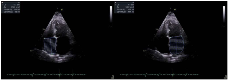
Standard 2D Echocardiography: From long axis pa-rasternal view, we measured the anteroposterior left atrium diameter, while we used the four-chamber view and two-chamber view to assess the left atrial (LA) area and volume (Figure 2). Area and volume va-lues were indexed to the body surface area (BSA).
Assessment of left atrial volume (LAV) was done in the apical four and two chamber views, using the Simpson biplane method. During a cardiac cycle, the following measurements were made: – maximal LA volume (LAV max) – measured at the end of ventricular systole, just before mitral valve opening (the end of the T wave of the ECG), indexed LAVI, and minimal LA volume (LAV min) measured at the end of diastole at mitral valve closure.
The same measurements were done for the right atrium (RA) – diameter, area and volumes obtained from apical four chamber view and afterwards we divided the volumes to the body surface area (RAVI – right atrium indexed volume) (Figure 3).
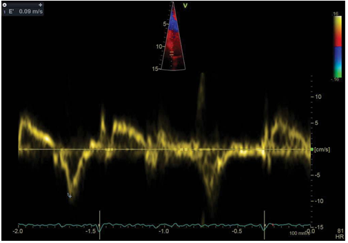
Filling pressures were estimated using the ratio between the peak early-diastolic transmitral flow ve-locity (E) and the average E’ (the mean between septal e’ and LV lateral e’) (Figure 4).
LA and RA phasic function was assessed using volu-metric parameters with previously measured volumes during the cardiac cycle.
The reservoir function – LA emptying fraction (LAEF) = (LAVmax-LAVmin)/LAVmax.
–RA emptying fraction (RAEF) = (RAVmax-RA-Vmin)/RAVmax.
For LA function index (LAFI) we measured in addi-tion the LV outflow tract velocity time integral (LVOT VTI – we used the mean on 3 consecutive beats) (Figure 5)8.
LAFI was calculated using the following formula7:
LAFI=(LAEF × LVOT)×VTI/LAVI
We used a similar method to calculate RA functional index (RAFI), using the measurement of VTI at the level of right ventricular outflow tract (the mean from 3 consecutive beats).
Statistical analysis
Measurements are presented as mean±SD. Variables were compared using Student’s t-test, ANOVA, or 2 test when appropriate. The relationships betwe-en different parameters were assessed by correlation analysis: Pearson’s method for continuous, normally distributed variables and Spearman’s rho method for ordinal or continuous but skewed variables. All statis-tical analyses were performed using SPSS 14.0 software for Windows (SPSS, Inc., Chicago, Illinois). A two-sided P-value of 0.05 was considered significant.

Figure 2 and 3. 2 dimensional echocardiography – LA and RA maximum volume in the apical 4 chamber view. LA – left atrium RA – right atrium.

Figure 4. Tissue Doppler interrogation of the septal wall at the level of the medial mitral annulus – measurement of the septal E’ wave (9 cm/s).

Figure 5. Pulsed wave doppler interrogation at the level of LVOT (Left ventricular outflow tract velocity time integral measure) – measurement of VTI (velocity time integral).
RESULTS
The demographic characteristics of the two groups are listed in Table 1. The mean age was 64.4 ± 9.6 years, 53% were women and mean BMI was 28.6 kg/ m2 ± 4.7 in all patients. These patients had at least one episode of atrial fibrillation.
P wave parameters in study groups
In patients with PAF, the maximum P wave duration was 99.4 ms ± 16.7; minimum P wave duration was 47.4 ms ± 9.8, the PD was 51.9 ms ± 12 and the P wave amplitude in DII was 0.99 ms ± 0.029. In the con-trol group, the same measurements were done and all are summarized in Table 1. In each of these instances, the difference between the 2 groups reached the level of significance (high significance for Pmin, Pd and Pa DII p<0.0001) and no significance for Pmax.

Echocardiographic atrial indices comparison Compared with the control group, patients with PAF had significantly larger anterior-posterior LA diameter, area and indexed volume. Patients with PAF had a sig-nifi cantly worse LA reservoir function (44.6 cm2±13.4 vs 61.4 cm2 ±9.4, p<0.0001) and LA functional index (26.8±11.6 cm2 vs 51.6±13.5 cm2, p<0.0001). In a simi-lar way, the PAF group had worse right atrial indices
– both the reservoir and the atrial function derived form RAFI were significantly altered compared with controls (47.1±13.6 vs 61.3±9.3 respectively 33±16.7 vs 51.5±13.5, p<0.0001). There were no significant differences between patients and controls regarding the RA volume and area, but patients with PAF had a larger RA mediolateral diameter.
Pd and echocardiographic atrial indices correlation
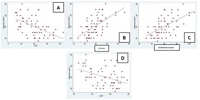
We used Pearson’s correlation to evaluate the relationship between Pd and echographic atrial parameters (Table 2) and we found a strong positive correlation with antero-posterior LA diameter, area and indexed volume (p<0.0001) and an inverse strong correlati-on with LA and RA function and index (p<0.0001). In addition, higher atrial electrical dispersion positively correlated with LV filling pressures. No correlation was found between Pd and RAVI, while Pd had a mo-derate positive correlation with RA area and mediola-teral RA diameter (Table 2).
DISCUSSIONS
Atrial fibrillation is associated with structural and elec-trical remodeling in the atria and ventricular myocar-dium. The heterogeneity in impulse conduction as a consequence of atrial fibrosis is one of the most im-portant electrical mechanism leading to AF. The progression of atrial fibrosis is the result of structural re-modeling and factors as a substrate for AF recurrence
- advanced atrial fibrosis is associated with increased risk for both paroxysmal and persistent or permanent AF. Our study aimed to evaluate the potential for simple and reproducible electrical parameters (P-wa-ve variability) derived from standard 12 leads ECG to potentially identify patients at risk of developing PAF and their correlation with echocardiographic atrial parameters to identify secondary atrial changes.
Several studies showed the relationship between P wave derived parameters and AF, like Gonna et al.9 who reported that parameters that accounted for the P-wave duration were increased in the recurrent AF than in the sinus rhythm group. P wave dispersion pro-ved to be the most specifi c and sensitive marker in distinguishing between the patients with PAF and nor-mal subjects. First time described by Dilaveris et al.10 in his study on 60 patients with PAF versus 40 healthy controls, the interlead variation in P wave duration is probably related to the inhomogeneity of electrical atrial activity that persists in patients with PAF even after restoring normal sinus rhythm. In our study Pd was significantly higher in patients with PAF compa-red to controls, these findings being consistent with previously reported data. For faster calculation and better reproducibility of Pd, automatic methods might be more accurate.
The correlation between Pd and echocardiographic left atrial indices such as left atrial size and function11, left atrial appendage function12 and the left ventricular diastolic function13,14 was described by several studies and was also demonstrated in our study. Another echocardiographic index evaluating the atrial function, independent of the underlying rhythm is the LA func-tion index (LAFI), a marker who incorporates surrogates of cardiac output, LA size and atrial reservoir function and which is inversely proportional to LA size and directly proportional to LA reservoir function and stroke volume.
Left atrial function index was associated with an increased risk of developing incident atrial fibrilla-tion independent of validated clinical risk prediction scores and echocardiographic measures of adverse cardiac remodeling. Left atrial function index can be easily measured using widely available 2-dimensional echocardiography and the studies demonstrated inde-pendent association of left atrial function index with adverse outcomes even in the presence of normal left atrial size, making it an enticing novel risk marker15. In our study, patients with PAF had worse LA mechanical function as assessed by LAFI compared to controls. Moreover, this is the fi rst study that established an inverse correlation between increased electrical dis-persion derived from Pd measurement on the ECG and LAFI/RAFI, underlying the link between impaired electrical activation of the atria and atrial mechanical dysfunction as assessed by both LAFI/RAFI and LAEF/ RAEF. We have also found a moderate correlation between Pd and LV fi lling pressures – probably beca-use the delay in electrical activation of the atria nega-tively impacts the diastole, especially if the left atrium activation is delayed, shortening the LV filling time and leading to a partially inefficient atrial contraction, conversely increasing LA pressure and in the end LV diastolic pressure.
Right atrium size is considered an independent pre-dictor of early PAF recurrence16 and a recent study by Luong C et al.17 on 95 patients with AF after cardio-version showed that right atrial volume is superior to left atrial volume for prediction of PAF. Considering these facts, we also analyzed the correlation betwe-en the electrical changes with RA dimensions and the parameters evaluating the function of the right atrium. To our knowledge, this is the first study that used RAFI to assess the performance of the right atrium, and we showed that patients with AF had poorer per-formance of the RA compared to controls.
In our study, echocardiographic atrial parameters (dimensions and function indices) and P wave para-meters were statistically different between the PAF group and the control group (patients with PAF had increased electrical dispersion and dimensions of LA/ RA, while the mechanical performance of RA/LA was worse in the PAF group), except P wave max. duration and RAVI – probably explained by the lack of specific and accurate measurement methods (automatic ECG measurement methods for Pd and Pmax, and 3 dimensi-onal echocardiography for a precise measurement of the right atrium) and the fact that at fi rst, RA is less involved in maintaining and initiating AF.
These indices are easy to asses in clinical practice and can help in evaluating the risk for developing AF in apparently healthy individuals, guiding the physician to screen these patients aggressively for AF, especially in the presence of symptoms (palpitations, thrombo-embolic events). Unfortunately, since we didn’t follow the patients prospectively, we cannot assess the pre-dictive value of these parameters for AF recurrence, which is one of the main limitations of our study.


Figure 6. Correlation between P wave dispersion (Pd) and left atrial indices.
A – correlation between Pd and left atrial function index. B – correlation between Pd and left atrial maximal area. C – correlation between Pd and left atrium maximal volume. D – correlation between Pd and left atrial emptying fraction.

Figure 7. Correlation between P wave dispersion (Pd) and right atrial indices.
A – correlation between Pd and right atrial emptying fraction. B – correlatin between Pd and right atrium mediolateral diameter. C – correlation between Pd and right atrial maximal area. D – correlation between Pd and right atrium maximal volume. E – correlation between Pd and right atrial function index.
LIMITATIONS
The small number of patients, the use of manual methods to measure P wave parameters, and the lack of long-term follow-up for patients in the PAF group, represent some of the limitations of this study.
CONCLUSIONS
Structural and electrical atrial remodeling is both a consequence and a substrate for atrial fibrillation. Be-cause of the high mortality in patients with untreated/ undetected AF, identifying patients at high risk for de-veloping AF is very important.
Easy to measure and reproducible markers are ne-eded in clinical practice for evaluating the risk of AF occurrence. Pd and atrial function indices proved to be meaningful in identifying susceptible patients, and mo-reover, electrical changes in atria are linked to mecha-nical disturbances, underlying the mechanism of atrial remodeling.
Conflict of interest: none declared.
References
1. Gialafos JE, Dilaveris PE, Gialafos EJ, Andrikopoulos GK, et al. P-wave dispersion: A valuable electrocardiographic marker for the prediction of paroxysmal lone atrial fibrillation. Ann Noninvas Elec-trocardiol 1999;4:39-45.
2. Ndrepepa G, Zrenner B, Deisenhofer I, Karch M, et al. Relationship between surface electrocardiogram characteristics and endocardial activation sequence in patients with typical atrial flutter. Z Kardiol 2000;89(6):527-37.
3. World Health Report 1998. Geneva: WHO, 1998, WA 540.1
4. Magnani JW, Mazzini MJ, Sullivan LM, et al. P-wave indices, distribu-tion and quality control assessment (from the Framingham Heart Study). Ann Noninvasive Electrocardiol 2010; 15: 77–84.
5. Aytemir K, Ozer N, Atalar E, et al. P wave dispersion on 12-lead electrocardiography in patients with paroxysmal atrial fibrillation. Pacing Clin Electrophysiol 2000;23(7):1109e12.
6. Lang RM, Badano LP, Mor-Avi V et al. Recommendations for cardiac chamber quantifi cation by echocardiography in adults: an update from the American Society of Echocardiography and the European Association of Cardiovascular Imaging. J Am Soc Echocardiogr. 2015; 28:1-39.e14.
7. Thomas L, Hoy M, Byth K, Schiller NB. The left atrial function in-dex: a rhythm independent marker of atrial function. Eur J Echocar-diogr. 2008; 9:356–362
8. Rosca M., Lancellotti P, Popescu BA, Piérard LA -Left atrial function: pathophysiology, echocardiographic assessment, and clinical applica-tions Heart. 2011 97(23):1982-9
9. Gonna H., Gallagher MM, Guo XH, Yap YG, Hnatkova K, Camm AJ. P-wave abnormality predicts recurrence of atrial fibrillation after electrical cardioversion: a prospective study. Ann Noninvasive Elec-trocardiol. 19, 57–62 (2014).
10. Dilaveris PE, Gialafos EJ, Sideris SK, et al. Simple electrocardiograph-ic markers for the prediction of paroxysmal idiopathic atrial fibrilla-tion. Am Heart J 1998; 135: 733–738
11. Tukek T, Akkaya V, Atilgan D, Demirel E, et al. Effect of left atrial size and function on P-wave dispersion: a study in patients with par-oxysmal atrial fibrillation. Clin Cardiol 2001; 24:676- 80.
12. Dogan A, Kahraman H, Ozturk M, Avsar A. P wave dispersion and left atrial appendage function for predicting recurrence after conver-sion of atrial fibrillation and relation of p wave dispersion to append-age function. Echocardiography 2004; 21:523-30.
13. Yilmaz R, Demirbag R, Durmus I, Kasap H, et al. Association of stage of left ventricular diastolic dysfunction with P wave dispersion and occurrence of atrial fibrillation after first acute anterior myocardial infarction. Ann Noninvasive Electrocardiol 2004; 9:330-8.
14. Gunduz H, Binak E, Arinc H, Akdemir R, et al. The relationship be-tween P wave dispersion and diastolic dysfunction. Tex Heart Inst J 2005;32:163-7.
15. Sardana M, Lessard D,Tsao C.V.; Parikh N.V.; Barton B; et al Asso-ciation of Left Atrial Function Index with Atrial Fibrillation and Car-diovascular Disease: The Framingham Offspring Study ;Journal of the American Heart Association 2018 Doi: 10.1161/Jaha.117.00843
16. Moon J, Hong YJ, Shim J, et al. Right atrial anatomical remodeling af-fects early outcomes of nonvalvular atrial fi brillation after radiofre-quency ablation. Circ J. 2012;76:860–867
17. Luong C, Thompson DJ, Bennett M, Gin K, Jue J, Barnes ME et al . Right atrial volume is superior to left atrial volume for prediction of atrial fibrillation recurrence after direct current cardioversion. Can J Cardiol. 2015 Jan;31(1):29-35.
 This work is licensed under a