Stefan Ailoaei1,2, Daniel Gafitescu1,2, Elisabeta Hurjui1, Elena Parvu3, Sorin Balasa4, Robert Alexandru5, Liviu Stoica2, Gabriela Creanga2, Mihaela Grecu1,2
1 Department of Cardiology, Institute of Cardiovascular Diseases „Prof. Dr. George I. M. Georgescu”, Iasi, Romania
2 Department of Electrophysiology, Institute of Cardiovascular Diseases “Prof. Dr. George I. M. Georgescu”, EHRA Recognized Training Center, Iasi, Romania
3 Hellimed SRL
4 Johnson&Johnson Company
5 Medtronic Company
Abstract: Electrophysiology is a rapidly evolving field of cardiology dedicated to the diagnosis and treatment of cardiac arrhythmias, with a wide range of procedures being performed in a complex environment. An interventional electrophysi-ology laboratory involves the acquisition of modern technological systems, and the presence of specialized and well trained medical personnel, consisting of at least one electrophysiologist, one technician and two nurses. To be in line with the current European standards, the laboratory must support the whole spectrum of interventions for heart rhythm disorders. In this regard, a powerful fluoroscopy system, programmable electrical stimulators and advanced ablation and 3D mapping systems are required. In recent years, the attention has focused on both the radiation dose minimization and protective radiological measures in the interventional cardiology laboratories, and thus the medical staff requires a specific set of qua-lifications. Continuous medical education is of paramount importance in an innovative field with such a steep technological rise as the field of electrophysiology. We considered it to be an opportune time to write this paper, as in February 2018 an official document attesting the training in electrophysiology and cardiac stimulation was officially launched by ministerial order in Romania.
Keywords: electrophysiology, training, education, laboratory, equipment
1. INTRODUCTION
Electrophysiology is a rapidly evolving field of cardio-logy dedicated to the diagnosis and treatment of cardi-ac arrhythmias, with a wide range of procedures being performed in a complex environment. The growing understanding of the mechanisms involved in the heart rhythm disorders and the continuous development of new modern technologies have granted electrophysio-logy studies the opportunity to be chosen as first line diagnostic and treatment options.
The electrophysiology laboratory
The electrophysiology laboratory is a special designed place to safely perform invasive procedures and must meet predefined standards in terms of space requi-rements and functionality in order to improve work conditions. The space allocated to the electrophysio-logy laboratory must be dynamic in order to facilitate the free movement of the staff in this environment, but at the same time to be able to accommodate all the equipment. The dimensions of the electrophysio-logy laboratory should be a minimum area of 33 m2, ideally 47 m2. Also, there should be a clear space of at least 2.43 m between the walls and the edges of the table where the patient lays. The height of the room depends on the size of the fluoroscopic equipment which can be mounted both in the fl oor or attached to the ceiling, the latter option allowing a more efficient cleaning of the floor2. There are special requirements regarding the facilities and equipment necessary for an electrophysiology laboratory, as well as the need for a well-trained staff. Among these facilities we mention the spaces for inpatient and outpatient care. The en-vironment in the electrophysiology laboratory should be sterile and should be provided with good air cir-culation, optimal brightness, ventilation and adequate heating /cooling3.
2. THE MEDICAL PERSONNEL
The unit dedicated to cardiac arrhythmia treatment involves not only state-of-the-art equipment and te-chnology but also well-trained personnel that must include minimum an electrophysiologist, two nurses and a technician. Thus, it is easy to infer that the staff working in the electrophysiology laboratory plays a central role in its proper functioning.
In most European countries, electrophysiology is not included among the compulsory modules of the residency program. Young fellows must train them-selves in accredited European centers which perform a wide and complete range of ablation and cardiac stimulation procedures in order to gain the neces-sary knowledge and competence1. Likewise, nurses working in the electrophysiology laboratory need additional knowledge regarding the diagnosis and treatment of cardiac arrhythmias. It is crucial to re-cognize the symptoms and clinical signs of arrhythmias and the various complications that may occur intra-or post-procedurally. Quick and effi cient management of these complications represents the key for both the patient safety and the better functioning of the electrophysiology laboratory. Furthermore, there is a special cardiovascular electrophysiology curriculum for nurses and there are accredited modules which provide comprehensive electrophysiology courses for those working in this field.
Electrophysiology training
The European Heart Rhythm Association (EHRA) pro-motes and seeks to ensure a homogeneous educati-on and qualification among European heart rhythm specialists, so it has organized a basic curriculum for arrhythmology specialists including the following: the Syllabus, the development of minimum standards and objectives for the electrophysiology training pro-gram (Curriculum), the creation of a model to cer-tify specialists in this fi eld and the training institutions (Accreditation) and the development of a registry for European electrophysiology specialists, their related institutions and their activity (Registries)4. Given the complexity of the mechanisms of different arrhythmi-as and their subsequent understanding, the duration of training in electrophysiology lasts for at least two years after finishing the residency in general cardio-logy. During these two years, the electrophysiology fellow must participate in the arrhythmology training program for at least 80% of the working hours (10% outpatient care, 10% device tracking, 10% device im-plantation, 40% invasive electrophysiology)5.
The Syllabus for the heart rhythm specialist is divided into two components. The first component consists in a general program that includes knowledge about anatomy, physiology, epidemiology, genetics and pathophysiology of rhythm disorders, arrhythmic di-seases and syndromes (e.g. ischemic and non-ischemic cardiomyopathy, channelopathies, hereditary syndro-mes) and techniques for the diagnosis and treatment of cardiac arrhythmias. The second component of the curriculum includes specifi c knowledge about invasive cardiac electrophysiology and implantable cardiac de-vices (equipment, principles, implantation / extraction and ablation techniques, complications). Therefore, the purpose of the basic curriculum is to determine the knowledge necessary for the trainee at the end of his/her training4,5. Moreover, the electrophysiology fellow must meet a minimal number of invasive and non-invasive procedures conducted as the first ope-rator. It is important to note that a European accre-dited training center must perform annually at least 250 invasive diagnostic procedures, 200 catheter abla-tion, 200 pacemaker implants/replacements, 50 ICD implants/replacements, and 20 CRT implants/replacements4. To date, in Romania there are only a few cen-ters that are able to meet this number of procedures, since the number of electrophysiologists in a given center is very small.
Obtaining European certification in electrophysiology
In order to obtain European certification in electro-physiology issued by the European Heart Rhythm Associ-ation, the fellow physician must undergo a proficiency examination that takes place once a year during the EHRA congress. The examination consists of a the-oretical part and in submitting a logbook within two years of the written exam which must contain at least 100 electrophysiological (self-standing or pre-ablation) electrophysiological studies and 100 catheter ablations as the first operator. The cases should be collected for two consecutive years4,6.
To date, there are 8 European accredited electro-physiologists in Romania, out of a total of 594 across Europe.
Obtaining national certification in electrophysiology
In February 2018, an official document attesting the training in electrophysiology and cardiac stimulation was officially launched in Romania by ministerial or-der, opening the perspective of national accreditation for young cardiologists who want to dedicate them-selves to the field of electrophysiology. The Electro-physiology and Cardiac Pacing training programs for the young cardiology specialists will be launched in the centers provided in the curriculum approved by the ministry, starting with the academic year 2018-2019.
Despite the strenuous, long way to reach the earli-est stages of the career as an electrophysiologist, even after obtaining a Free Practice Certificate, continuo-us medical education remains the cornerstone of this profession and regular educational programs are im-perative for maintaining a high level of competence in line with the international standards1.
Maintaining competence in electrophysiology
Continuous Medical Education (EMC) in all subspecial-ties of cardiology is essential, since the knowledge and skills needed in these areas are in permanent develop-ment. Standards are maintained through continuous learning and practice, as major changes can occur at any time in this field. Thus, it is necessary to participa-te annually in at least two international seminars and international conferences. Continuous medical prac-tice requires at least 16 work hours per week in the field of electrophysiology, while continuous medical education is defi ned by the accumulation of 200 EMC credits over a five-year period. The Accreditation as an electrophysiologist is valid for 10 years, after which a new process of reaccreditation and recertification is necessary4.
3. EQUIPMENT
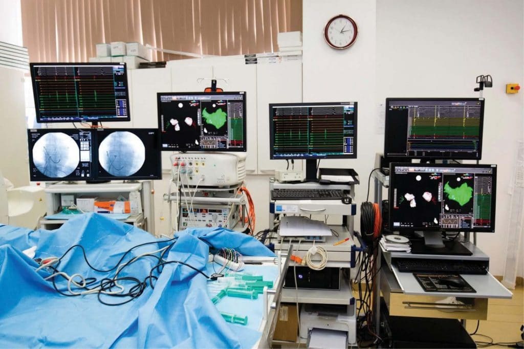
The standard requirements of an electrophysiology laboratory include a state-of-the-art fluoroscopy system, programmable electrical stimulators, integra-ted data display systems, 3D mapping systems that are used for the ablation of complex arrhythmias such as atrial fi brillation or ventricular tachycardia, as well as catheter ablation systems designed to fit the anatomy of each patient (Figure 1). There are several systems for recording the surface electrocardiogram and the intracardiac electrograms, depending on the purcha-se price, the quality of the signals provided and the maintenance costs. The minimum requirements for a monitoring system should include a 12-lead surface electrocardiogram and a 24-channel intracardiac elec-trogram. For the more advanced laboratories such as those able to perform complex ablations, there are systems with up to 64-128 intracardiac channels. The electrophysiologist represents the decisive factor in choosing a work system, as he has to be familiar with it and with the installation of the latest software1,7.
Electroanatomic 3D mapping systems used for real-time reconstruction of each patient’s heart ana-tomy are especially required for complex ablations, such as pulmonary vein isolation or ablation of ven-tricular tachycardia. There are three mapping systems available on the market: CARTO 3D from Biosense Webster Inc. and Ensite NavX at St. Jude Medical Inc and Rhythmia HDxTM from Boston Scientific Corpo-ration. The CARTO 3D system is based on obtaining the catheter’s virtual position using a static magnetic field, while the Ensite NavX system depends on the constant measurement of impedance. Robotic naviga-tion systems such as Stereotaxis™ and Electromecha-nics (Sensei™; Hansen Medical) facilitate mapping and ablation of complex arrhythmias, both offering the advantage of being able to manipulate the catheters inside a control room, thus substantially reducing the exposure of the staff to radiation8,9.
Many laboratories that perform complex ablation procedures have digital fluoroscopic systems with advanced imaging capabilities such as rotational angio-graphy, rotational CT imaging and offer modalities to integrate 3D CT and MRI imaging. Reconstructed 3D imaging based on CT or cardiac MRI helps the electro-physiologist to guide the catheter and choose an abla-tion technique. A common case is represented by the atrial fibrillation ablation procedure when a preproce-dural 3D image may prove to be extremely useful if the patient presents particularities in the anatomy of the left atrium and / or the pulmonary veins10,11.
The programmable electrical stimulators are the cornerstone in conducting an electrophysiological study and have between two to four programmable output channels in order to adjust the amplitude and the duration of the stimulus. Burst pacing and extrasti-muli testing coupled at different cycle lengths are stan-dard in the electrophysiology laboratory, being able to reveal the mechanism of an arrhythmia and to diffe-rentiate between intracardiac potentials, or to simply measure the refractory period of the tested structu-res7.
In order to perform a safe catheter ablation pro-cedure, an electrophysiology laboratory requires a modern ablation system which includes generators, cables and catheters connected to an energy source in the form of radiofrequency, cryo, ultrasound, or laser ablation. The most widely used sources are ra-diofrequency and cryo ablation. Over the years, radi-ofrequency ablation has been proven to be the most effective and safe therapeutic method for the treat-ment of a wide range of cardiac arrhythmias. How-ever, the selection of a particular ablation technique depends on the operator’s preference, the anatomical particularities of the patient and, last but not least, the ablation target12,13.
Radiation
The field of interventional cardiac electrophysiology is dependent on images obtained by the fluoroscopy system in order to position catheters/electrodes cor-rectly within the heart. Thus, the exposure of the me-dical staff and of the patient to radiation is inevitable. Radiation protection and minimization to the exposu-re have become priorities in interventional cardiology laboratories, therefore the medical staff must be well-trained in the field of radiation safety7.
The most important parameters recorded by the fluoroscopic equipment are the fluoroscopic time, the radiation dose (expressed in Gy) which measures the deterministic effects of the potential injury, and the dose-area product (cGy cm2) which measures the stochastic effects of the potential injury. Deterministic effects refer to harmful tissue reactions caused by a threshold dose of absorbed radiation (e.g. skin burn), whereas stochastic effects include malignant prolifera-tion due to DNA damage. The latter are not directly determined by the radiation dose, but higher doses in-crease the likelihood of adverse effects. It is important to note that the stochastic effects of repeated exposu-re to radiation are cumulative, and the physician who exposes the patient to fluoroscopy should primarily consider the irradiating history of the patient14-16.
It is intuitive to conclude that in order to minimize the patient’s exposure to radiation, it is needed to re-duce the radiation dose as much as possible. The most effective factors contributing to the reduction of ra-diation exposure are the minimization of fluoroscopy time and the decrease of the fluoroscopy pulse rate, at the cost of temporal resolution of the image. The main measures required in order to reduce the exposure to radiation of the medical staff are increasing the dis-tance to the source of radiation, minimizing the scatte-red radiation and limiting the dose used. In this regard, a protective apron, eye protection, thyroid protection and proper table shielding are of paramount importan-ce for greatly reducing the radiation. Moreover, the distance from the radiation source considerably redu-ces the dose received, as it decreases with the square of the distance from the source17,18.
Pre-procedural care
The pre-procedural preparation of the patient should be performed by qualified medical personnel, as the correct positioning of all the patches, the correct re-cording of the surface electrocardiogram, the adminis-tration of the necessary medication, the careful moni-toring of the vital parameters and the ease in manipula-ting the systems’ hardware and software are necessary skills for the proper functioning of the laboratory1,7. Most electrophysiological studies are performed after the discontinuation of the antiarrhythmic drug thera-py. The procedures that involve the left chambers of the heart are performed under oral anticoagulation and / or intra-procedural intravenous anticoagulation. To the greatest extent, the procedures are performed under local anesthesia. Sometimes profound anesthe-sia is required, especially for complex ablations such as atrial fibrillation, ventricular tachycardia or pediatric ablation. Orotracheal intubation is preferred in the case of ventricular tachycardia ablation, particularly in electrical storms, as hemodynamic instability may be high and there is often a need for multiple intra-pro-cedural external electrical shocks.
Post-procedural monitoring
Patients should be carefully monitored electrocardio-graphically and hemodynamically in a recovery or in-tensive care unit after complex ablation procedures and implantation of devices. In this regard, external cardiac defi brillators, pacemakers, and transthora-cic echocardiography should be available at all times. Immediate echocardiography, as well as a day after the procedure should be performed in all patients in or-der to exclude the presence of pericardial effusion. Moreover, patients who have benefited from the im-plantation of a cardiac device should routinely perform a chest X-ray to exclude pneumothorax and to evalu-ate the position of the electrodes within the heart. In cases where pseudo-aneurysms, arteriovenous fistula, or groin hematoma is suspected at the site of punctu-re, duplex ultrasonography should be performed. Last but not least, it is important to note that a close colla-boration between a large volume electrophysiology center and a cardiac surgery unit represents an essen-tial step that provides backup in case of cardiovascular emergencies1,7.
The outpatient clinic
The outpatient clinic consists of two components: a unit designed for the treatment of cardiac tachyar-rhythmias and another for bradyarrhythmias and im-plantation of pacemakers. In most cases, patients are directed to the electrophysiology center by other physicians within the territory, but there are cases when they are self-referrals and are evaluated on the basis of the symptoms and on the interpretation of the electrocardiogram that documents the arrhythmia.
The arrhythmological consultation involves explain-ing treatment options, including interventional treat-ment when the patient has indication. An integral part of the consultation is represented by the pre-sentation of the hospitalization time, the risks of the procedure and the success rates according to the electrophysiologist’s own experience. A 12-lead elec-trocardiogram, echocardiography, Holter monitoring, and exercise-testing are immediately available to the patient, while computer tomography, coronary angi-ography and nuclear magnetic resonance imaging may be scheduled if necessary.
All patients that undergo an ablation or implant procedure are consulted in the outpatient clinic at least one day prior to admission and informed con-sent is obtained prior to each procedure. Usually, the vast majority of the patients can be discharged one day after the procedure, after receiving a letter that contains the procedure protocol, the physician’s re-commendations, and a scheduled date for the next consultation.
Another key component of the electrophysiology program is represented by the postprocedural follow-up of all patients in order to assess their current status, to analyze their heart rate and rhythm, to interrogate their cardiac device, or to discuss other treatment op-tions. Hospitals that offer outpatient monitoring have the advantage of being able to track the evolution of the patient in the long term, thus improving the quality of the medical service. New telemonitoring technolo-gies are gaining more and more ground by providing the possibility of immediate transmission of recorded data. Therefore, in the case of an arrhythmically li-fe-threatening event, the on-call physician can mana-ge the situation in a very short time, increasing the patient’s chances of survival. Moreover, regular visits to the doctor may also no longer be necessary in pa-tients without arrhythmic events or, on the contrary, they may be promptly arranged in patients with acute arrhythmic symptoms or events1.
Conflict of interest: none declared.
References
1. Kuck, K., Wissner, E. and Metzner, A. (2012). How to Establish an Arrhythmia Unit in the 21st Century. Revista Española de Cardio-logía (English Edition), 65(1), pp.92-96.
2. Facilities Guidelines Institute. Guidelines for design and construction of health care facilities. http://www.fgiguidelines.org/. Published Janu-ary 1, 2014.
3. Zipes, D., Calkins, H., Daubert, J., Ellenbogen, K., Field, M., Fisher, J., Fogel, R., Frankel, D., Gupta, A., Indik, J., Kusumoto, F., Lindsay, B., Marine, J., Mehta, L., Mendes, L., Miller, J., Munger, T., Sauer, W., Shen, W., Stevenson, W., Su, W., Tracy, C. and Tsiperfal, A. (2015). 2015 ACC/AHA/HRS Advanced Training Statement on Clinical Car-diac Electrophysiology (A Revision of the ACC/AHA 2006 Update of the Clinical Competence Statement on Invasive Electrophysiol-ogy Studies, Catheter Ablation, and Cardioversion). Circulation: Ar-rhythmia and Electrophysiology, p.HAE.0000000000000014
4. Merino JL, Arribas F, Botto GL, Huikuri H, Kraemer LI, Linde C, et al. 2005-2007 Accreditation Committee of the European Heart Rhythm Association. Core curriculum for the heart rhythm special-ist. Europace. 2009;11(Suppl 3):iii1-iii26.
5. Kearney P, Oktay Ergene A, Escaned J, Flachskmpf F, Griebenow R, Kristesen S et al. Education Committee of the European Society of Cardiology. Core Curriculum for the General Cardiologist. Nice, France: European Society of Cardiology; 2006.
6. Escardio.org. (2018). Invasive Cardiac Electrophysiology Certifi ca-tion. [online] Available at: https://www.escardio.org/Education/Ca-reer-Development/Certifi cation/Invasive-cardiac-electrophysiology.
7. Haines, D., Beheiry, S., Akar, J., Baker, J., Beinborn, D., Beshai, J., Brysiewicz, N., Chiu-Man, C., Collins, K., Dare, M., Fetterly, K., Fish-er, J., Hongo, R., Irefin, S., Lopez, J., Miller, J., Perry, J., Slotwiner, D., Tomassoni, G. and Weiss, E. (2014). Heart Rhythm Society Ex-pert Consensus Statement on Electrophysiology Laboratory Stan-dards: Process, Protocols, Equipment, Personnel, and Safety. Heart Rhythm, 11(8), pp.e9-e51.
8. Chun KR, Wissner E, Koektuerk B, Konstantinidou M, Schmidt B, Zerm T, et al. Remote-controlled magnetic pulmonary vein isola-tion using a new irrigated-tip catheter in patients with atrial fibrilla-tion. Circ Arrhythm Electrophysiol. 2010;3:458-64.
9. Schmidt B, Tilz RR, Neven K, Julian Chun KR, Fürnkranz A, Ouyang F. Remote robotic navigation and electroanatomical mapping for ab-lation of atrial fibrillation: considerations for navigation and impact on procedural outcome. Circ Arrhythm Electrophysiol. 2009;2:120-8.
10. Gupta S, Desjardins B, Baman T, Ilg K, Good E, Crawford T, Oral H, Pelosi F, Chugh A, Morady F, Bogun F, Delayed-enhanced MR. scar imaging and intraprocedural registration into an electroanatomical mapping system in post- infarction patients. JACC Cardiovasc Imag-ing 2012;5(2):207-210.
11. Haqqani HM, Tschabrunn CM, Tzou WS, et al. Isolated septal sub-strate for ventricular tachycardia in nonischemic dilated cardiomy-opathy: incidence, characterization, and implications. Heart Rhythm 2011;8(8):1169-1176.
12. Scheinman M, Calkins H, Gillette P, Klein R, Lerman BB, Morady F, Saksena S, Waldo A. NASPE policy statement on catheter ablation: personnel, policy, procedures, and therapeutic recommendations. Pacing Clin Electrophysiol 2003;26(3):789-799.
13. Chanani NK, Chiesa NA, Dubin AM, Avasarala K, Van Hare GF, Col-lins KK. Cryoablation for atrioventricular nodal reentrant tachycar-dia in young patients: predictors of recurrence. Pacing Clin Electro-physiol 2008;31(9):1152-1159.
14. Vano E, Rosenstein M, Liniecki J, Rehani MM, Martin CJ, Vetter RJ. ICRP Publication 113. Education and training in radiological pro-tection for diagnostic and interventional procedures. Ann ICRP 2009;39(5):7-68.
15. Gerber TC, Carr JJ, Arai AE, et al. Ionizing radiation in cardiac imag-ing: a science advisory from the American Heart Association Com-mittee on Cardiac Imaging of the Council on Clinical Cardiology and Committee on Cardiovas- cular Imaging and Intervention of the Council on Cardiovascular Radiology and Intervention. Circulation 2009;119(7):1056-1065.
16. Glazier JJ, Dixon SR. Skin injury following prolonged fluoroscopy: early and late appearances. QJM 2012;105(6):571-573.
17. Hirshfeld JW Jr, Balter S, Brinker JA, et al. ACCF/AHA/HRS/SCAI clinical competence statement on physician knowledge to optimize patient safety and image quality in fluoroscopically guided invasive cardiovascular procedures. A report of the American College of Cardiology Foundation/American Heart Association/American Col-lege of Physicians Task Force on Clinical Compe- tence and Training. J Am Coll Cardiol 2004;44(11):2259-2282.
18. Einstein AJ, Moser KW, Thompson RC, et al. Radiation dose to pa-tients from cardiac diagnostic imaging. Circulation 2007;116(11): 1290-1305.
 This work is licensed under a