Ileana Arsenescu
Article received on the 21st May 2012. Article accepted on the 28th May 2012.
1 „Prof. Dr. C. C. Iliescu” Emergency Institute for Cardiovascular Diseases, Bucharest
„Prof. Dr. C. C. Iliescu” Emergency Institute for Cardiovascular Diseases, Bucharest
28 year old patient, clinically presenting a right lateral-cervical tumoral formation.
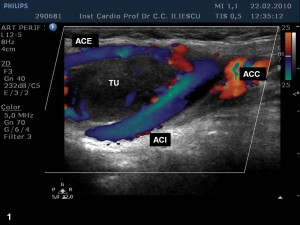
Figure 1. 2D and color ecography shows a mass (TU) situated cranial to the carotid bifurcation, between the internal and external carotid arteries. It is well – defined with a hypoechogenic, homogeneous structure.
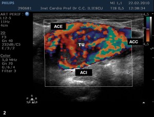
Figure 2. Decreasing Doppler speed scale, we note that the described mass is highly vascularized, with slow flow speeds.
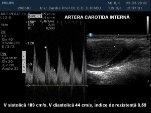
Figure 3. At spectral Doppler exam, speed and the resistance indices are normal on the internal carotid artery.
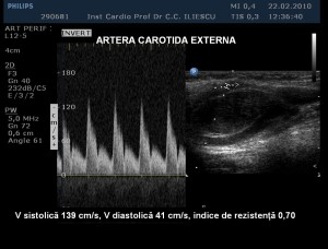
Figure 4. At the level of the external carotid artery, systolic velocities are higher, indicating an increased flow. The resistance index is low due to the increase in the diastolic velocity, showing that the artery is irrigating a low resistance area, that is the glomic tumor.
 This work is licensed under a