Dragos Cozma1,2, Cristina Vacarescu2, Simina Crisan1,2, Emilia Violeta Goanta2, Mihai Andrei Lazar2, Cristian Mornos1,2, Adina Ionac1,2, Lucian Petrescu1,2
1 “Victor Babes” University of Medicine and Pharmacy, Timisoara, Romania
2 Institute of Cardiovascular Diseases, Timisoara, Romania
We report the case of a 65-year-old woman that recently presented with heart failure NYHA class III symptoms (intermittent nocturnal dyspnea, dizziness, asthenia) and postero-thoracic discomfort and pain. The patient, diagnosed with permanent atrial fibrillation since 1999, underwent mitral valve replacement surgery (for severe mitral regurgitation due to anterior mitral leaflet prolapse) as well as atrial septal defect closure and tricuspid annuloplasty in 2001. At that time, an 11.5 cm antero-posterior diameter of the left atrium (LA) was reported from the parasternal long
axis view; no other data are available regarding LA volume or surface. At the present, approximately 14 years after surgery, a giant LA of 1451 ml was measured by transthoracic echocardiography (four chamber view, area-length method). The mitral valve bi-leaflet prosthesis Orbis 31 was normally functioning and the left ventricular (LV) EF was 32% (LV end diastolic volume of 167 ml). To our surprise, a clear window to the entire heart was only available by left postero-thoracic view (while searching for pleural effusion, which was absent). Dense LA spontaneous echo contrast and a large LA appendage (volume 24.97 ml) were also clearly documented, but no clinical thromboembolic events were noted. Symptoms of low cardiac output due to left ventricular dysfunction were associated with symptoms of pulmonary congestion due to severe left atrium enlargement and elevated left ventricular fi lling pressure, also the patient had severe thorax skeletal changes with scoliosis and respiratory function impairment. Open chest surgery for LA volume reduction was proposed but the patient refused the intervention and was discharged after symptomatic
improvement on medical treatment with ACE inhibitors, beta-blocker, diuretics and anticoagulation. Giant left atrium (GLA) is a rare condition characterized by huge enlargement of the left atrium with a diameter exceeding 65 mm, with a reported incidence of 0.3 %1,2 and is commonly associated with longstanding rheumatic mitral valve regurgitation. Not many cases of “non-rheumatic” GLA are available but its identification is essential because of the complications associated: atrial fibrillation, thromboembolic complications, hemodynamic and sever respiratory complications3. Volume reduction is useful to prevent complications; the indication for surgery is the presence of intracardiac or extracardiac compressive symptoms and several techniques have been proposed such as simple wall placation or resection and primary suture which is more effective and more secure in volume reduction4,5. Although investigations such as transesophageal ecochardiography or computer tomography could better describe left atrium morphology and intrathoracic relations, transthoracic echography provides the primary diagnosis and in such cases a different approach could be necessary for a complete assessment. Conflict of interests: none declared.
References:
1. Ahmad K Darwazah, Hamdy El Sayed Giant left atrium associated with massive thrombus formation, Thrombosis Journal 2013, DOI: 10.1186/1477-9560-11-5.
2. Ahmed El Maghraby, Rachel Hajar – Giant Left Atrium: A Review, Heart Views. 2012 Apr-Jun; 13(2): 46-52.
3. Imran Ahmed, Achyut Sarkar, Arindam Pande, and Chanchal Kundu – A “non-rheumatic” giant left atrium, Ann Pediatr Cardiol. 2015 Jan- Apr; 8(1): 95-97.
4. Tamura Y, Nagasaka S, Abe T, Taniguchi S – Reasonable and effective volume reduction of a giant left atrium associated with mitral valve disease. Ann Thorac Cardiovasc Surg. 2008 Aug;14(4):252-5.
5. Funk M1, Perez M, Santana O. Asymptomatic giant left atrium. Clin Cardiol. 2010 Jun;33(6):E104-5. doi: 10.1002/clc.20736.
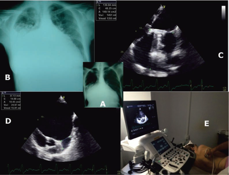
A. Chest x-ray postero-anterior view demonstrate markedly enlarged cardiac silhouette. There is a double contour to the right heart border and splaying of the carina. There are no overt features of congestive cardiac failure at this time. (Year 2001)
B. Chest x-ray postero-anterior view demonstrate also markedly cardiomegaly with signs of pulmonary edema (year 2014).
C. Giant LA of 1451 ml measured by transthoracic echocardiography (four chamber view, area-length method).
D. E. Left postero-thoracic view for a clear window to the entire heart.
 This work is licensed under a