Oana Ionita1,2, Eva Paskova2, Robert Petr1,2, Lucie Srncova2, Hana Linkova1,2
1 3rd Faculty of Medicine, Charles University, Prague, Czech Republic
2 IIIrd Clinic of Internal Medicine and Cardiology, Cardiocenter, University Hospital Královské Vinohrady, Prague, Czech Republic
Abstract: Free floating thrombi in the right atrium are a rare entity and they are most frequently described in patients with pulmonary embolism1. In 1989 the European Working Group on Echocardiography identified three morphological patterns of right heart thrombi2: worm-shaped, highly mobile thrombi arising from the lower limb veins (type A thrombi), non-mobile, nonspecific clots that resemble left heart thrombi and are most likely formed in situ (type B thrombi) and a more rare type of clots which are highly mobile but not wormlike (type C). Overall prognosis in patients with right heart thrombi is dismal, however significant differences in the potential of embolization and short-term mortality based on the morphology of the clot are encountered2.
Keywords: right atrial thrombus
We report the case of an 89-year-old patient admitted for mild left hemiparesis. CT showed ischemic changes in the right parietal regions with no signs of hemorr-hage, NIH stroke scale score was 4 and conservative treatment for stroke was initiated. Patient denied any history of dyspnea, angina or syncope. Physical exami-nation was unremarkable except for the hemiparesis and irregular heart rhythm.
ECG showed atrial fibrillation (first documentation) and nonspecific ST-T changes. Carotid Doppler ultra-sonography showed no significant stenosis.
During hospitalization a transthoracic echocardi-ography was performed, which revealed normal left ventricular systolic function and fibrotic changes of the mitral and aortic valves with mild regurgitations. In the right atrium a big, highly mobile echogenic mass was noted, with no attachment to the right atrial structu-res (Figure 1 and 2). The mass measured approxima-tely 23×28 mm and showed chaotic displacement in the right atrium, being occasionally trapped in a „ping-pong“ like movement among the tricuspid valve and the right atrial ceiling (Figure 3). Right ventricle had normal dimensions but reduced longitudinal function. No interatrial shunt was detected transthoracically.
The characteristics of the mass were consistent with a right atrial thrombus. Patient refused further imaging examinations (transesophageal echocardio-graphy, chest CT), so no data on possible coexistent left atrial thrombus or interatrial communication was available. He also refused any non-conservative treatment option. Oral anticoagulation was started and the patient was discharged home with complete remission of neurologic signs.
A couple of months later transthoracic echocardio-graphy showed complete resolution of the right atrial thrombus under anticoagulation therapy. The pati-ent had a good functional status and no clinical event suggesting pulmonary embolism was identified.
Compared with type A and type B thrombi in the right atrium, type C thrombi have an intermediate risk of embolization and thrombus-related complica-tions. In our case of a type C thrombus, conservative treatment was chosen based on the patient´s wish and advanced age.
Despite the echocardiographic appearance sugges-ting high risk of embolization, the clinical outcome with anticoagulation therapy was favorable.
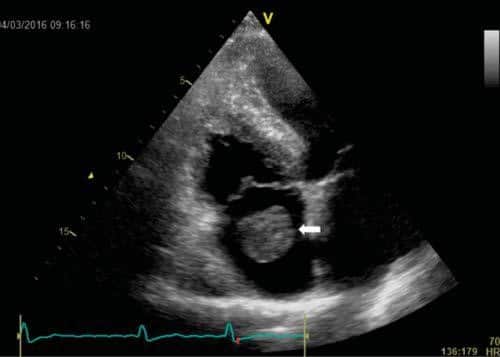
Figure 1. Transthoracic echocardiography, apical 4-chamber view focused on the right cavities showing big ball-like thrombus in the right atrium (ar-row) with no attachment to the right atrial structures.
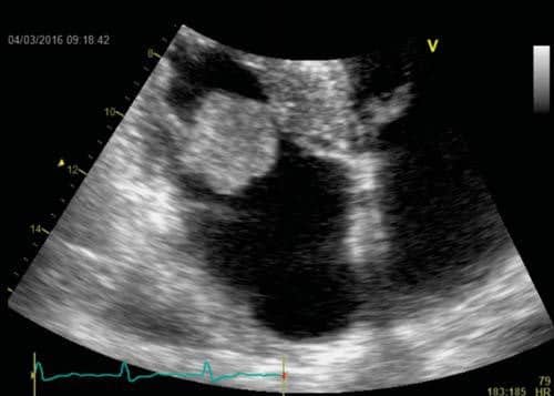
Figure 2. Apical 4-chamber view – zoom on the right atrium. The throm-bus intermittently protrudes through the tricuspid valve.
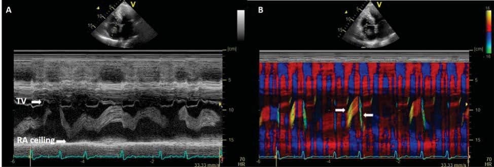
Figure 3. A. M-mode through the right heart chambers showing extreme mobility of the mass between the planes of tricuspid valve (TV) and the atrial ceiling (arrows). B. Superimposed tissue velocity imaging shows intermittent changes of the velocity of the thrombus (arrows).
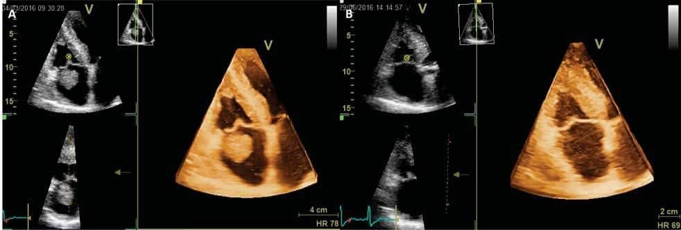
Figure 4. 3D transthoracic echocardiography volume data – acquisition from apical 4-chamber view – at the initial presentation (A) and two months later (B) showing complete disappearance of thrombus.
Conflict of interests: none declared.
References
1. Chartier L, Béra J, Delomez M, Asseman P, Beregi JP, Bauchart JJ, Warembourg H , Théry C. Free-Floating Thrombi in the Right Heart. Diagnosis, Management, and Prognostic Indexes in 38 Conse-cutive Patients. Circulation. 1999;99:2779-2783.
2. Kronik G. The European Cooperative Study on the clinical signifi-cance of right heart thrombi. European Heart Journal (1989) 10, 1046-1059.
 This work is licensed under a