Download PDF
https://doi.org/10.47803/rjc.2020.30.1.56
Adriana Ioana Ardelean1,2, Madalina Ioana Moisi1,2, Sabina Belenes2, Marius Rus1,3, Mircea Ioachim Popescu1,3
1 Department of Preclinical Disciplines, Faculty of Medicine and Pharmacy, University of Oradea, Oradea, Romania
2 Emergency Clinical County Hospital of Oradea, Oradea, Romania
3 Department of Clinical Disciplines, Faculty of Medicine and Pharmacy, University of Oradea, Oradea, Romania
Abstract: Myocardial infarction with nonobstructive coronary arteries disease (MINOCA) is defi ned as a clinical syndro-me with normal or near normal coronary arteries disease (stenosis severity revealed on angiography less than 50%), exclu-ding other overt causes of myocardial ischemia such as cardiac trauma. Trials report an incidence between 5-10% among subjects with acute myocardial infarction (AMI). The optimal management and the outcome of this syndrome require the proper identification of the specific pathophysiologic mechanism. We report the case of a 67-year-old male presenting with the clinical and enzymatic picture of a myocardial infarction with ST segment elevation in the anterior territory with no significant coronary arteries stenosis on the angiography. Our case illustrates the importance of revealing the real underlying condition which is involved in producing the MINOCA syndrome, because this is the only strategy suitable for a correct treatment and a favorable outcome.
Keywords: MINOCA, angiography, coronary artery stenosis, antiplatelet therapy.
Rezumat: Infarctul miocardic cu artere coronare permeabile (MINOCA) este definit ca un sindrom clinic cu artere coronare normale sau aproape normale (definite prin severitatea stenozei coronariene mai mică de 50%, decelată cu ocazia coronarografiei), excluzând alte cauze de ischemie miocardică, cum ar fi traumatismele cardiace. Studiile au raportat o incidenţă între 5-10% în cazul subiecţilor cu infarct miocardic acut (AMI). Managementul optim şi prognosticul acestui sindrom necesită identificarea corespunzătoare a mecanismului fi ziopatologic specifi c. Raportăm cazul unui bărbat în vârstă 76 de ani care prezintă tabloul clinic şi enzimatic a unui infarct miocardic cu supradenivelare de segment ST în teritoriul anterior, fără stenoze coronariane semnificativă evidenţiate prin coronarografie. Cazul nostru ilustrează importanţa dezvăluirii cauzei reale incriminate în producerea sindromului MINOCA, deoarece aceasta este singura strategie potrivită pentru un tratament corect şi o evoluţie favorabilă.
Cuvinte-cheie: MINOCA, angiografi e, stenoză coronariană, terapie antiplachetară.
INTRODUCTION
Atherosclerosis represents the main process which may lead to the occlusion of the epicardial arteries in ST-segment-elevation myocardial infarction (STEMI). Usually the presence of the obstructive coronary artery disease is revealed in 95% of the cases with STEMI and NON-STEMI1.
The intriguing MINOCA syndrome was underdiagnosed in the past because clinicians thought that the absence of the obstructive atheromatous plaque or the acute thrombosis represents the exclusion argu-ments of a true ST-segment-elevation myocardial in-farction, leading to a misinformation and assurance of a favorable outcome in these subjects. The whole idea was redefined when the Fourth Universal Defi nition of the Myocardial Infarction was released in 2018. MI-NOCA syndrome is defi ned as the presence of the acute myocardial infarction without signifi cant coronary artery disease on the angiography. The diagnosis should exclude other overt causes that might cause troponin elevation, like sepsis or pulmonary embo-lism, Takotsubo syndrome or myocardial cell injury that is not related to ischemia (eg: myocarditis)2.
The Vigo study revealed an incidence of MINOCA syndrome of more than 10% in young patients with acute myocardial infarction. Even if the clinical parti-cularities of the MINOCA subjects differs comparing with the AMI group, the mortality rate at 1 month and 1 year are similar proving that the myocardial infarcti-on with nonobstructive coronary artery disease is not a benign condition and further tests should reveal the underlying etiology1.
There are some mechanisms that might be involved in the appearance of the MINOCA syndrome inclu-ding the atheromatous plaque erosion, embolism or epicardial coronary artery dissection. Other causes refer to a vasospasm of a coronary artery or of the microcirculation.
Besides angiography, the etiological diagnosis could be provided by the echocardiography, intravascular ultrasound, optical coherence tomography2. Cardiac magnetic resonance represents an important exami-nation that may reveal late gadolinium enhancement caused by tissue damage due to myocardial infarction3.
Coronary CT angiography may be used for iden-tifying of the atherosclerotic marks, with the ob-servation that plaque rupture or erosion cannot be illustrated using this method. Description of normal coronary artery on coronary CT angiography does not guarantee the absence of a thrombotic disease as a main underlying condition for MINOCA occurrence. This is the reason why the diagnosis should be formulated only if the absence of a stenotic lesion is revealed using the coronary angiography rather than considering the presence of atherosclerosis landmarks described by noninvasive imaging elements for etiolo-gical diagnosis of MINOCA3.
CASE PRESENTATION
We report the case of a 67-year-old male, without previous medical history, presenting in the emergency department with chest pain, sweating, accompanied by a new left bundle branch block on the electrocardio-graphy (Figure 1). High sensitivity troponin illustrated a high value (5187 ng/L) and the others biological fi n-dings were in normal ranges, except from an eleva-ted blood glucose (263 mg/dl) and high glycosylated hemoglobin (8.8%). The echocardiography reflected global hypokinesia of the left ventricle and a severe reduction of ejection fraction (25%) (Figure 2). Emer-gency angiography was performed using transradial approach but there were no significant coronary arte-ries stenosis, with an estimated TIMI flow 3 in all the epicardial coronary arteries (Figure 3 and 4).
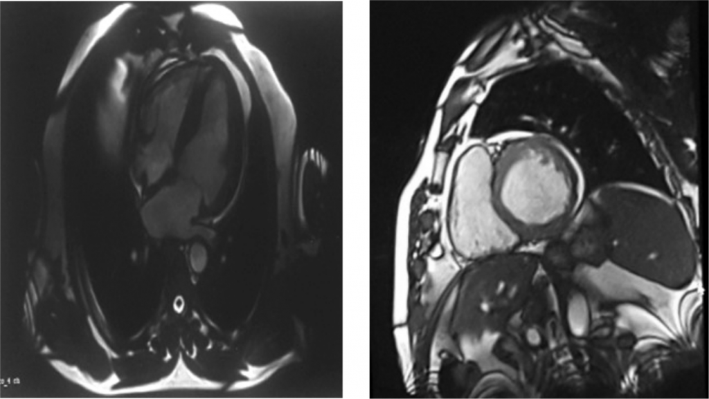
Considering the electrocardiographic aspect, corro-borated with the elevation of the myocardial necrosis markers and the angiography, we decided that the di-agnosis was MINOCA syndrome, which required furt-her investigations. The diagnostic algorithm was com-pleted by cardiac magnetic resonance, an important diagnostic method, which revealed necrosis of the la-teral ventricular wall, with no edema or microvascular obstructions. The dilated left ventricle illustrated a de-pressed ejection fraction on the CMR (43%), with an indexed myocardial mass in the normal ranges. Finally, the conclusion emphasized a transmural lateral myo-cardial infarction in the circumfl ex coronary artery territory, with no myocardial cells viability (Figure 5).
Analyzing the CMR result and the angiography wi-thout relevant coronary arteries stenosis, the con-clusion was that exist a certain underlying substrate which is producing a predisposition for thrombosis. We finally performed the genetic tests for detection of the thrombophilia and the result was surprising. The complex profile described a combination of hete-rozygous and homozygous mutations of the elements involved in the coagulation cascade, predisposing to a prothrombotic status. The heterozygous mutations were found for the V H1299R (R2) factor, II G20210A factor, XII V34L factor and PAI-1 4G/5G. In additi-on, there was a homozygous mutation of the MTHFR C677T, revealing a high predisposition for arterial and venous thrombosis.
In our opinion that the main underlying cause was an arterial thrombus even if the coronary angiography did not reveal this aspect, this would be the suitable explanations consistent with the cardiac MRI descrip-tion. In situ formation and subsequent spontaneous lysis of a coronary artery thrombus represents a plau-sible mechanism involved in MINOCA syndrome.
The most challenging aspect regarding this case is represented by the identification of a proper treat-ment, considering the fact that the patient has the pre-disposition for both arterial and venous thrombosis and the existing trials are still debating whether there is certain indication of the direct oral anticoagulants (DOACs). Our option was antivitamin K drugs due to the fact that there are clinical trials with strong evi-dences regarding their reduction of thrombotic events in subjects with thrombophilia. Besides the oral anti-coagulants, the treatment included antiplatelet thera-py, diuretics, inhibitors of the angiotensin enzyme and insulin.

Figure 1. Electrocardiogram performed at admission reflects sinus rhythm, 87 beats/minute, left bundle branch block.


Figure 2. Transthoracic echocardiography: parasternal long axis (right) and apical 5 chambers (left) – reflects dilatation of the left ventricle.

Figure 3. Angiography – Left and circumflex coronary arteries with no obstructive lesions.

Figure 4. Angiography – Right coronary artery without significant stenosis.

Figure 5. Cardiac MRI – vertical – long axis (left) and mid ventricular short axis (right) revealing lateral myocardial infarction due to late gadolinium en-hancement caused by cellular necrosis.
DISCUSSIONS
MINOCA syndrome should be faced as a working di-agnosis and all the efforts should be directed towards finding the real underlying substrate. The outcome of this syndrome represents a serious problem, with an estimated one year mortality of 4.7%4.
Investigations like cardiac magnetic resonance and coronary imaging accompanied by functional assess-ment are required for the etiological diagnosis of MI-NOCA.
An observational study involving MINOCA patients from the SWEDEHEART registry demonstrated that the reinfarction rate in these subjects was 6% during a mean follow-up of 4.3 years. The mortality rate was equally high after recurrent MINOCA, as compared to the myocardial infarction with significant coronary artery stenosis with an initial MINOCA episode. This idea reflects the fact that MINOCA is not a benign condition and patients who repeat the event will have a negative outcome5.
Another relevant registry, named VIRGO, expre-ssed the fact that MINOCA is not a harmless conditi-on, revealing that a similar number of both MINOCA and AMI with coronary artery disease experienced heart failure and cardiac arrest6.
Clear indications regarding the dual antiplatelet the-rapy in MINOCA are lacking. The MINOCA registry gathered 9.466 subjects without etiological diagnosis of this syndrome. This registry aimed to discover the optimal medical therapy for secondary prevention and the long-term outcome. The conclusion of this regis-try was that there is a long term benefit in using the ACE inhibitors or angiotensin II receptor blockers and a positive effect of the beta-blockers, but the dual an-tiplatelet therapy expressed a neutral effect4.
Data from randomized trials should be used to identify the optimal management of MINOCA, with a clear benefit on the secondary prevention.
The association between antiphospholipid syndro-me (APS) and MINOCA incidence was revealed in an observational study. MINOCA rate had a higher incidence in subjects with antiphospholipid syndrome compared with the control group without APS7.
The indication of DOACs in MINOCA syndrome caused by thrombophilia remains controversial. The-re is only one trial which compared rivaroxaban with warfarin for secondary prevention of venous thrombo-embolism in patients with APS but the evidences were not strong enough to favors the usage of DOACs7.
Arterial thrombosis in patients with MINOCA and APS has indication of antivitamin K, but there is a chal-lenge in maintaining the INR between the recommen-ded ranges because the APS may produce false elevation of the INR. The recurrent arterial thrombotic events despite a therapeutic INR may be solved by adding aspirin, statins or using a low-molecular-weight heparin8.
CONCLUSIONS
The etiological diagnosis of the MINOCA syndrome represents the essential key in fi nding the proper ma-nagement of this condition. CMR had clear benefi ts in revealing the underlying cause of the MINOCA syndrome in our patient and gave us the clue to per-form the genetic tests for thrombophilia.
Further randomized studies are required to esta-blish whether the DOACs represent a better option than antivitamin K in patients with MINOCA and APS. We considered that antivitamin K agents are the best therapeutic approach in this particular case, but the thrombotic event may repeat because there is still a challenge to maintain the INR in therapeutic ranges and offer the best antithrombotic protection.
Conflict of interest: none declared.
Referennces:
1. Pasupathy S., Tavella R., Beltrame J.F. Myocardial Infarction With Nonobstructive Coronary Arteries (MINOCA) The Past, Present, and Future Management. Circulation. 2017;135:1490–1493.
2. Thygesen K., Alpert J.S., Jaffe A.S., Chaitman B.R. et al. Fourth uni-versal defi nition of myocardial infarction. European Heart Journal. 2018, 40(3): 237–269.
3. Agewall S., Beltrame J.F., Reynolds H.R., Niessner A. et al. ESC work-ing group position paper on myocardial infarction with non-obstruc-tive coronary arteries, European Heart Journal, 2017, 38(3):143– 153.
4. Lindahl B, Baron T, Erlinge D, Hadziosmanovic N et al. Medical ther-apy for secondary prevention and longterm outcome in patients with myocardial infarction with nonobstructive coronary artery disease. Circulation. 2017;135:1481–1489.
5. Nordenskjold AM, Baron T, Eggers KM, Jernberg T, Lindahl B. Pre-dictors of adverse outcome in patients with myocardial infarction with non-obstructive coronary artery (MINOCA) disease. Int J Car-diol. 2018;261:18–23.
6. Kochanek KD, Murphy SL, Xu J, Tejada-Vera B. Deaths: final data for 2014. Natl Vital Stat Rep. 2016;65:1–122.
7. Gandhi H., Ahmed N., Spevack D.M. Prevalence of myocardi-al infarction with non-obstructive coronary arteries (MINOCA) amongst acute coronary syndrome in patients with antiphospholipid syndrome. IJC Heart & Vasculature. 2019,22:148–149.
8. Garcia D., Erkan D. Diagnosis and Management of the Antiphospho-lipid Syndrome. N Engl J Med 378;21:2010-2020.
 This work is licensed under a