Elena Bivol1, Livi Grib1, Alexandra Grejdieru1
1 „Nicolae Testemiţanu” State University of Medicine and Pharmacy, Chisinau, Republic of Moldavia
Abstract: Cardiovascular disease prevalence is steadily increasing leading to high mortality and morbidity. The diagnosis of cardiorenal syndrome is a real challenge. Objectives – Estimation of glomerular filtration rate in cardiorenal syndrome diagnosis in patients with heart failure with reduced and mid-range ejection fraction. Methods – The prospective study included 170 patients with reduced and mid-range ejection fraction heart failure (HF) hospitalized during January 2016 – December 2017 period in the Cardiology Unit of Municipal Clinical Hospital „Holy Trinity” in Chisinau. Results – A total of 170 patients were evaluated: 83 subjects with chronic HF and cardiorenal syndrome (CRS) and 87 subjects with chronic HF without renal involvement. The estimated glomerular filtration rate (eGFR) estimated by means of EPI equation based on cystatin C and creatinine level had a mean of 43.40 ± 1.29 ml/min/1.73 m2 in cardiorenal syndrome group and a mean of 78.29 ± 1.34 – ≤60 mL/min/1.73 m2 in the group without kidney involvement. Conclusions – Study results confirm the superiority of GFR estimation by means of EPI equations based on cystatin C and creatinine level for cardiorenal syndrome screening and early diagnosis.
Keywords: cardiorenal syndrome, heart failure, glomerular filtration rate, cystatin C, creatinine.
INTRODUCTION
International defi nitions and agreements regarding sta-ging of acute, chronic kidney disease and renal injury are under constant review. In 2002, NKF-KDOQI1 proposed a staging model for CKD (chronic renal di-sease) based on five estimated glomerular filtration rate (eGFR) intervals. Subsequently, the model was approved by KDIGO (Kidney Disease Improving Global Outcomes) with some changes. CKD was defi ned as an eGFR <60 ml/min/1.73 m2, this threshold representing half of the normal GFR for young adults. Additionally, the threshold represents the point value at which the-re is an increase in prevalence and severity of several cardiovascular risk factors and in CKD specific labo-ratory changes2. The applicability of KDIGO criteria in CKD and renal involvement definition in patients with HF has already been proven for an eGFR threshold 60 ml/min/1.73 m2. This critical point was used in a series of HF studies3 and registries4 in order to assess significant renal involvement and prognostic value in relation to morbidity, mortality, and re-hospitalizati-on rate. Glomerular filtration rate (GFR) is internationally recognized as the best overall kidney index5. Although existing equations are adjusted for several parameters that infl uence creatinine level, such as age, gender and race, GFR estimates should be interpre-ted with caution6. A further concern is validation of equations in test populations. These issues should be taken into account when assessing renal involvement in HF, considering that most participants are over 65 years old7 with GFR decreasing gradually with age. The MDRD formula is validated in this population, althou-gh CKD-EPI is more accurate in some cases8.
Purpose of the study: Estimation of glomerular fil-tration rate in cardiorenal syndrome diagnosis in pa-tients with heart failure with reduced and mid-range ejection fraction.
Material and methods: The prospective study in-cluded 170 patients with heart failure (HF) with redu-ced and mid-range ejection fraction, 83 subjects with type 2 cardiorenal syndrome (CRS) and 87 subjects with no renal involvement, hospitalized during January 2016 – December 2017 period in the Cardiology Unit of Municipal Clinical Hospital „Holy Trinity” in Chisi-nau, Republic of Moldova.
The type 2 CRS was diagnosed according to the working group of the 11th Conference of the Consen-sus ADQI (2013)9,10.
Inclusion criteria:
CHF diagnosis (as defined in the 2016 ESC Gui-delines for the diagnosis and treatment of acute and chronic heart failure)11. LVEF ≤49%.
No previous renal impairment (CKD) or docu-mented onset of heart failure prior to renal im-pairment onset.
Age over 18 years.
Exclusion criteria:
Presence of primary kidney disease (congenital kidney disease or kidney disorders prior to car-diac disease).
Inflammatory, traumatic kidney disease (that can-not be explained by CHF).
Steroid treatment.
Patients with acute cardiac/cerebrovascular events.
Arterial hypertension was defined according to 2018 ESC/ESH Guidelines for the management of arterial hypertension criteria (sistolic BP ≥140 mmHg and/or diastolic BP≥90 mmHg)12.
Standard echographic views (Hitachi HI VISION Avius device) using the Simpson biplane method ad-justed for frame rate optimization were obtained to measure chamber dimensions and evaluate global and regional left ventricular function.
NT-proBNP (N-terminal pro-B-type natriuretic pepti-de) – was assessed using electrochemiluminescence immunoassay (ECLIA). Cutt-off value for non- acute patients was considered 125 pg/ml.
Creatinine – was determined through kinetic method (Siemens Dimension EXL 200). Cutt-off value: in male <1.2 mg/dL, in female <1.0 mg/dL.
Cistatine-C – determinated through the nephelometric method. Cutt-off value: under 50 y.o.: 0.55-1.15 mg/l; older than 50 y.o.: 0.63-1.44 mg/l. BUN – determinated through the spectrophotometric method.
Evaluation of renal function aimed at assessing es-timated glomerular fi ltration rate (eGFR) according to previously described formulas and degree of renal involvement according to K/DOQI classifi cation: renal involvement with normal GFR (G1) ≥90 ml/min/1.73 m2; renal involvement with slightly reduced GFR (G2): 60-89 ml/min/1.73 m2; renal involvement with mode-rate GFR decrease (G3a): 45-59 ml/min/1.73 m2 and GFR (G3b): 45-59 ml/min/1.73 m2; severe GFR decre-ase (G4): 15-29 ml/min/1.73 m2; end-stage renal disea-se (ESRD) with GFR (G5) <15 ml/min/1.73 m2.
We assessed GFR using CKD-EPI equation based on cystatin C and creatinine levels (GFR reference va-lues ≤60 ml/min/1.73 m2) in order to evaluate CRS prevalence, divide study groups, and perform compa-rative analysis.
CKD-EPI equation based on cystatin C and crea-tinine levels (ml/min/1.73 m2) = GFRcyscr = 135 × min (SCr/ , 1) × max (SCr/ , 1) – 0.601 × min (Scys/0.8, 1) – 0.375 × max (Scys/0.8, 1) – 0.711 × 0.995 Age × 0.969 [in women] × 1.08 [in African American race], where Scr is serum creatinine, is 0.7 for women and 0.9 for men, is –0.248 for women and –0.207 for men, min indicates the minimal value for SCr/ or 1, max indicates the maximal value for SCr/ or 1, and Scys is serum cystatin C.
Additionally, GFR was determined by other formulas:
CKD-EPI equation based on creatinine level GFRepi (ml/min/1.73 m2) = 141 x min (SCr/k, 1) x max (SCr/k, 1) – 1.209 x 0.993 age x 1.018 [for
women] x 1.159 [for African American race], where SCr is serum creatinine level (mg/dL), k is 0.7 for women and 0.9 for men, is –0.329 for women and –0.411 for men, min is the minimal value of SCr/k or 1 and max is the maximal value of SCr/k or 1.
CKD-EPI equation based on cystatin C level (ml/ min/1.73 m2) = GFRcys = 133 x min (Scys/0.8, 1)– 0.499 x max (Scys/0.8, 1) – 1.328 x 0.996 age x 0.932 [for women], where Scys is serum cystatin C.
Short MDRD equation (Modification of Diet în Renal Disease) based on 4 variables: GFRm (ml/ min/1.73 m2) = 186 x (serum creatinine μmol/l x 0,0113) – 1,154 x age (years) – 0.203 x 1. 212 (for African American race) x 0,742 (for women);
Classical Cockroft Gault equation: GFRCG cre-atinine clearance (ml/min/1.73 m2) = [140 – age] x weight (kg) x 1.23 (for men) or x 1.04 (for wo-men)/serum creatinine μmol/l.
Simple equation based on cystatin C: 100/cystatin C (mg/l); GFR100/cys (ml/min).
It is more prudent to take into account the result of the patient’s actual GFR, not the GFR result that the patient could have if his BSA were 1.73 m2. Indexation of GFR for BSA can induce relevant differences in pa-tients with abnormal body size.
GFR estimation equations are adjusted to standard body surface area (1.73 m2). We checked the diagnos-tic value of unadjusted and adjusted to body surface area equations. Unadjusted eGFR was assessed by MDRD, CKD-EPI based on creatinine, CKD-EPI ba-sed on cystatin C and CKD-EPI based on cystatin C and creatinine: eGFR (ml/min) x BSA (m2)/1.73, where body surface area BSA (m2) = (W 0.425 x H 0.725) x 0.007184.
ETHICAL ASPECTS
The participants were informed about the subject, purpose and rules of the study. Each participant signed and agreed on admission to participate in the research process, their data being processed anonymously.
STATISTICAL ANALYSIS
All data was statistically analyzed using the SPSS v20 for Windows, Microsoft Excel for Windows 10 software, univariate statistical analysis (frequency, mean, range, and median) and comparison test being performed variables Chi2, Student’s t test comparing two means (quantitative). Data was expressed as mean ± SD, whi-le for p-value we were using the two-tailed test.
RESULTS
We determined a mean GFRcyscr value of 43.40 ± 1.29 ml/min/1.73 m2 (95% CI 40.82-45.98, p <0.05) for the study group with variations ranging from 14 to 59 ml/ min/1.73 m2. The control group had a mean eGFR of 78.29 ± 1.34 (95% CI 75.63-80.94, p <0.05), ranging from 60 to 113 ml/min/1.73 m2 (Figure. 1).
According to K/DOQI stages of renal involvement we identified G1 stage in 25.29% cases and G2 stage in 74.71% cases, comprising the control group; G3a stage in 54.9% cases; G3b in 29.3% cases; G4 in 14.6% cases and G5 in 1.2% (1 patient), comprising the study group.
Table 1 represents mean eGFR values calculated by different equations. Comparative analysis determined a high variability among obtained values: ranging from 37.77 ml/min/m2 by CKD-EPI equation based on cystatin C to 70.37 ml/min/m2 by Cockroft Gault classical equation for the study group; and from 67.64 ml/min/ m2 by CKD-EPI equation based on cystatin C to 125.6 ml/min/m2 by Cockroft Gault equation for the control group. The extreme values in both groups were obtai-ned using these two formulas, whereas the results obtained using the unadjusted cystatin C CKD-EPI equation had the closest values compared to CKD-EPI equation results based on cystatin C and creatinine (formula used for sample division): 44.33 mL/min/1.73 m2 vs. 43.39 ml/min/1.73 m2 in the study group and 79.22 vs. 78.29 ml/min/1.73 m2 in the control group. In our study, equations based only on serum creatinine overestimated GFR values compared to serum cysta-tin C equations (Table 1).
We examined ROC curves for GFR estimation equations in order to assess diagnostic value of each test relative to GFRcyscr. Ideally, the effi cacy of a GFR estimation test should be compared to GFR measured by plasma and urinary clearance of exogenous mar-kers. Inulin is an ideal exogenous marker, but alterna-tive markers such as iothalamate, EDTA, DTPA and iohexol can be used as well. Unfortunately, measu-rement of exogenous markers clearance is complex, expensive and difficult to apply in routine clinical prac-tice.
The ROC (Receiver Operating Characteristics) curve is a bidimensional curve where Y axis indicates sensiti-vity while X axis indicates specificity. The curve helps us measure a model’s efficiency. The higher the area under the curve (maximum is 1), the better the mo-del. Analyzing data in Table 1, the AUC (area under the ROC curve) has maximal values for GFRcys (0.94) and GFRepi (0.92) equations compared to GFRcyscr, whi-le having a minimal value (0.87) for Cockroft Gault equation. Maximum sensitivity was assessed for GFRepi (84.34%) and GFRcys (84.15%), but for the optimal di-fferent criterion GFR ≤ 73ml/min/m2 vs. GFR ≤ 50 ml/min/m2. GFR100-cys showed maximum specificity (89.39%), however the smallest sensitivity (78.05%) relative to GFRcyscr. The maximum positive predicti-ve value was established for GFR100-cys (73.9%) and GFRcys (73.8%), while the lowest positive predictive value (59.3%) was found for eGFR based on classi-cal Cockroft Gault equation. Absence of renal invol-vement was most accurately appreciated by cystatin C based eGFR (negative predictive value -93.7%, p<0.001) and GFRepi (negative predictive value -93.6%, p <0.001).
When comparing adjusted and unadjusted for body surface area GFR estimation methods (Figures 2-8), we notice an increased efficiency for adjusted equa-tions: 0.906 vs. 0.901 for GFRm and GFRmG respecti-vely; 0.915 vs. 0.905 for GFRepi and GFRepiG; 0.936 vs. 0.928 for GFRcys and GFRcysG. The difference may also be due to equation selection to which the adjusted GFRcyscr was calculated.
Table 1 data and Figures 2-8 demonstrate that all examined models are effective for renal involvement diagnosis in HF patients. All estimation methods pro-ved to be excellent diagnostic models except for the classical Cockroft Gault equation estimation method (AUC 0.87, p <0.001) that was appreciated as a good estimation model from sustained data having a value analysis p = 0.0001. The maximal diagnostic value was established for unadjusted CKD-EPI equations based on cystatin and creatinine levels, and adjusted CKD-EPI based on cystatin and creatinine.
We analyzed age influence on GFR, dividing patients into groups using the 65-year delimitation threshold (Figure 9).
We obtained the following results: for the group with no renal involvement GFRcyscr was 81.02 ± 1.82 ml/min/m2 in the below 65-year age group and 74.42 ± 1.77 ml/min/m2 in the ≥65-year group (p <0.05). The study group had GFRcyscr values of 44.48 ± 2.18 ml/ min/m2 in the below 65-year age group and 42.88 ± 1.62 ml/min/m2 in the ≥65-year group (p>0.05). For both groups, GFRcyscr was elevated in advanced age subjects.
Subsequently, we assessed hypertension influence on eGFR level. (Fig. 10)
The recorded level of GFRcyscr in patients with CRS had lower values in case of hypertension association with values of 42.76 ± 1.48 ml/min/m2 vs. 46.27 ± 2.48 ml/min/m2 in the absence of hypertension (p <0.05). No statistically significant differences were found in the control group: 78.35 ± 1.63 ml/min/m2 for hypertensive subjects vs. 78.10 ± 2.19 ml/min/m2 in the ab-sence of hypertension (p > 0.05). The phenomenon, however, is not due solely to hypertension presence but also to coexisting factors – left ventricular ejection fraction (LVEF) in control group was lower in patients without hypertension 37% vs. 41.16% (p <0.01) and NT proBNP levels were significantly higher in non-hypertensive subjects 4712.05 pg/dl vs. 1974.17 pg/dl (p <0.001), suggesting a more serious evolution of HF in this group.
Next, we evaluated serum cystatin level. Mean cystatin C value was 1.79 ± 0.06 mg/dl (95% CI 1.67-1.91) for patients with CRS and 1.12 ± 0.02 mg/dl (95% CI 1.09-1.19) in those without CRS (p <0.001). Test sensitivity was 78.05% while specificity was 89.39% for the optimal creatinine >1.38 mg/dl criterion, with the positive predictive value -90.2% and negative predicti-ve value -76.4%.
Further, we evaluated serum creatinine levels in our groups. Mean creatinine value was 1.38 ± 0.07 mg/dl (95% CI 1.24-1.52) for patients with CRS and 0.79 ± 0.02 mg/dl (95% CI 0.75-0.84) in those without CRS (p <0.001). Test sensitivity was 78.05% while specificity was 86.36% for the optimal creatinine >0.92 mg/dl le-vel, positive predictive value being 87.8% and negative predictive value 75.8%.
Serum BUN level was 10.88 ± 0.54 mmol/l in pa-tients with impaired renal function and 8.28 ± 0.28 mmol/l in those with a GFR >60 ml/min/m2 (p <0.001).
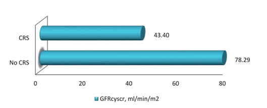
Figure 1. GFRcyscr value in patients with/without CRS.

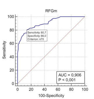
Figure 2. ROC curve for MDRD equation.
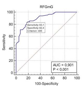
Figure 3. ROC curve for unadjusted MDRD equation.
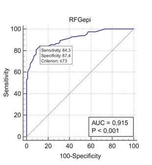
Figure 4. ROC curve for creatinine based CKD-EPI equation.
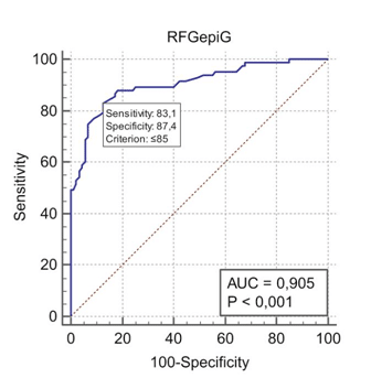
Figure 5. ROC curve for unadjusted creatinine based CKD-EPI equation.
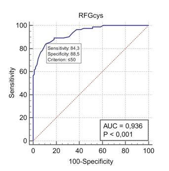
Figure 6. ROC curve for cystatin based CKD-EPI equation.
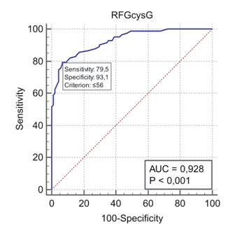
Figure 7. ROC curve for unadjusted cystatin based CKD-EPI equation.
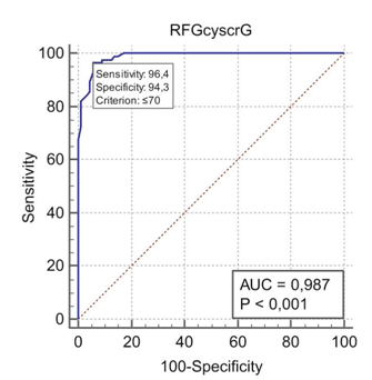
Figure 8. ROC curve for unadjusted cystatin and creatinine based CKD-EPI equation.
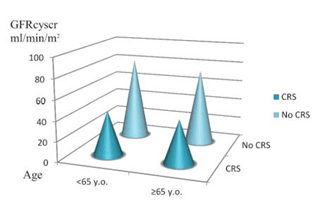
Figure 9. Patients’ distribution according to age groups.

Figure 10. GFRcyscr level according to hypertension and CRS presence.
DISCUSSIONS
Improving cardiorenal risk assessment and stratificati-on is crucial. In real life, measuring GFR with an exo-genous marker is rarely possible. Thus, we continue relying on GFR assessment by endogenous markers such as creatinine and cystatin C. We usually clas-sify patients according to eGFR making no difference among molecules used for measurement. Basically, we assume that we will have the same result for any of the markers.
In order to highlight the difference between GFR estimation methods in clinical practice (other than AUC), we will exemplify. If we assume that we have a cohort of 170 subjects comprised of all participants from our study, not dividing them into two different study groups, CRS rate assessed by cystatin and cre-atinine based CKD-EPI equation will be 48.82%, by unadjusted cystatin and creatinine based CKD-EPI equation will be 32.94%, by cystatin based CKD-EPI equation will be 66.47%, by unadjusted cystatin based CKD-EPI equation will be 48.24%, by creatinine ba-sed CKD-EPI equation will be 28.82%, by unadjusted creatinine based CKD-EPI equation will be 21.18%, by MDRD equation will be 30.59%, by unadjusted MDRD equation will be 22.94%, by cystatin-100 based equa-tion will be 36.47% and by Cockroft Gault classical equation will be 20.59% (p <0.001).
This phenomenon was also described by Swedish researcher Åkerblom in 2015 who performed a uni-centric observational study involving outpatient cardiac patients (n = 2716), cardiology unit patients (n = 980), coronary heart disease unit patients (n = 1464). He attempted to reclassify patients with non-acute cardiac disease distributed inaccurately according to eGFRepi compared to eGFRcys. Differences are more evident in more critical situations when we need fir-mer decisions, however Åkerblom demonstrated that only 53 out of 143 patients with GFRcys could be di-agnosed with GFRepi, whereas in 8 cases signifi cant kidney involvement was excluded13. Overestimations of creatinine-based GFR were recorded in all groups, with a mean of 10 ml/min/m2 at a GFR level <90 ml/ min/m2. Several studies have assessed the delayed cre-atinine elevation in acute cardiac pathology, so Swe-dish researchers split outpatient and cardiology unit patients in groups without acute pathology. In pati-ents with GFRcys <30 ml/min/m2, GFRepi had a 13 ml/ min/m2 (22 vs. 35 ml/min/m2) higher level; for GFRcys = 30 – 59 ml/min/m2, GFRepi had a 16 ml/min/m2 (44 vs. 60 ml/min/m2) higher level; for GFRcys = 60 – 89 ml/ min/m2, GFRepi had a 10 ml/min/m2 (74 vs. 84 ml/min/m2) higher level and for GFR ≥90 ml/min/m2, there was a 1 ml/min/m2 difference (101 vs. 102 ml/min/m2). Another study performed by Kervella D et al. com-pared GFR levels assessed by different methods in a cohort of subjects with type 2 CRS. Their results con-firm GFR overestimation using GFRepi equation (41 ± 20mL/min/1.73 m2) compared to GFRcys (30 ± 15 mL/ min/1.73 m2), GFRcyscr (34 ± 15 mL/min/1.73 m2) or GFRm (26 ± 11 mL/min/1.73 m2)14.
In conclusion, GFR estimation in HF patients will be more accurate when using adjusted/unadjusted CKD-EPI equations based on cystatin and creatinine levels, adjusted cystatin based CKD-EPI and adjusted creati-nine based CKD-EPI.
Analyzing the ROC curve in order to assess cystatin C in CRS diagnostic value, we obtained an AUC of 0.9 (95% CI 0.84-0.94, p <0.001), cystatin C level being an excellent model for CRS diagnosis, but less accurate compared to eGFR. Cystatin C levels can be affected by thyroid dysfunction or steroid use, and can have reduced specificity in concomitant infections15, situ-ations that cannot be totally excluded in hospitalized patients.
Existing studies assess cystatin C as a marker for early differential diagnosis of acute renal injury, and as a prognostic parameter in these patients16. As a routine kidney biomarker, compared to serum cre-atinine, cystatin C is disadvantaged by higher costs, limited accessibility and number of specialists famili-ar with its reference values and limited use in GFR estimation formulas17. Serum creatinine has remained a „gold standard” in clinical practice, being affordable and cost-effective, with medical staff being familiar with its interpretation, and having evidence to support its use in clinical setting. Studies evaluating worsening renal function impact in HF are not an exception to this approach18,19.
When examining the ROC curve for assessing cre-atinine diagnostic value in CRS, we obtained an AUC value of 0.877 (95% CI 0.81-0.93, p <0.001). In this way, although creatinine level is a good model for CRS diagnosis, it is less efficient compared to cystatin C and GFR estimated by most equations, except for the Cockroft Gault equation with similar diagnostic value (AUC-0.87).
Serum BUN level was higher in patients with im-paired renal function. Similar data were obtained by Palazzuoli21 who performed an analysis of a cohort of 246 subjects or by Salim20 who investigated a cohort of 563 subjects with or without HF either associated or not with renal changes.
Traditional renal biomarkers do not provide in-formation regarding the level or cause of kidney dys-function. Creatinine may be infl uenced by food intake, muscle mass, gender, medication and other disorders, having, in addition, a slow response compared to other renal biomarkers. Serum BUN level can be infl uenced by hepatic dysfunction, gastrointestinal hemorrhages, dehydration, steroid use, or protein intake3.
CONCLUSIONS
- GFR assessment plays a key role in cardiorenal syndrome diagnosis. Compared to GFRcyscr, ma-ximum diagnostic value was found for GFRcys (AUC ROC 0.94) and GFRepi (AUC ROC 0.92) equations, maximal sensitivity was determined for GFRepi (84.34%) and GFRcys (84.15%), while GFR100-cys had maximum specificity (89.39%).
- GFR estimation using the EPI equation based on cystatin C level or based on cystatin C and crea-tinine level could be used for the purpose of CRS screening or early diagnosis.
- Glomerular filtration rate estimation using the classical Cockroft Gault equation showed mini-mal diagnostic value (AUC ROC 0.87) and the lowest positive predictive value (59.3%).
Conflict of interest: none declared.
References
1. Levey, A.S., Coresh, J., Balk, E., Kausz, A.T., Levin, A., Steffes, M.W. et al „National Kidney Foundation practice guidelines for chronic kidney disease: evaluation, classification, and stratification.” National Kidney. 2003;139(7):605.
2. Sarnak M.J., Levey A.S., Schoolwerth A.C., Coresh J., Culleton B., Hamm L.L. et al „Kidney Disease as a Risk Factor for Development of Cardiovascular Disease: A Statement From the American Heart Association Councils on Kidney in Cardiovascular Disease, High Blood Pressure Research, Clinical Cardiology, and Epidemiology and Prevention” Circulation. 2003; 108:2154–2169.
3. Silva, P., Nikitin, N.P., Witte, K.K.A., Rigby, A.S., Loh, H. „Effects of applying a standardised management algorithm for moderate to se-vere renal dysfunction in patients with chronic stable heart failure.” European Journal of Heart Failure. 2007; 9:415–423. ISSN: 1879-0844.0.
4. Heywood, J.T., Fonarow, G.C., Yancy, C.W., Albert, N.M., Curtis, A.B., Stough, W.G. et al. „Influence of renal function on the use of guideline-recommended therapies for patients with heart failure.” Am J Cardiol., 2010. 105(8):1140-6., ISSN: 0002-9149.
5. Eknoyan G., Levin N.W. Impact of the New K/DOQI Guidelines. Vols. Blood Purif 2002;20:103–108. https://doi.org/10.1159/000046992,
6. Michels WM1, Grootendorst DC, Verduijn M, Elliott EG, Dekker FW, Krediet RT. Performance of the Cockcroft-Gault, MDRD, and new CKD-EPI formulas in relation to GFR, age, and body size. Vols. Clin J Am Soc Nephrol. 2010 Jun;5(6):1003-9. doi: 10.2215/ CJN.06870909.
7. Rácz O., Lepej J., Fodor B., Lepejová K., Jarčuška P., Kováčová A. and The Hepameta Study Group. Pitfalls in the Measurements and Asses-ment of Glomerular Filtration Rate and How to Escape them. Vols. EJIFCC. 2012 Jul; 23(2): 33–40.
8. McAlister FA1, Ezekowitz J, Tarantini L, Squire I, Komajda M, Bayes-Genis A, Gotsman I, Whalley G, Earle N, Poppe KK, Doughty RN and Investigators., Meta-analysis Global Group in Chronic Heart Failure (MAGGIC). Renal dysfunction in patients with heart failure with preserved versus reduced ejection fraction: impact of the new Chronic Kidney Disease-Epidemiology Collaboration Group for-mula. Vols. Circ Heart Fail. 2012 May 1;5(3):309-14. doi: 10.1161/ Circheartfailure.111.966242.
9. Cruz, D.N., Schmidt-Ott, K.M., Vescovo, G., House, A.A., Kellum, J.A., Ronco, C., Mccullough, P.A. „Pathophysiology of Cardiorenal Syndrome Type 2 în Stable Chronic Heart Failure: Workgroup State-ments from the Eleventh Consensus Conference of the Acute Dialy-sis Quality Initiative (ADQI).” Contrib Nephrol., 2013; 182:117-36. ISSN:1662-2782.
10. Rangaswami, J., Bhalla, V., Blair, J.E.A. et. al. „Cardiorenal Syndrome: Classification, Pathophysiology, Diagnosis, and Treatment Strategies: A Scientifi c Statement From the American Heart Association”. Cir-culation. 2019 March; 139(16):e840-e878. https://doi.org/10.1161/ CIR.0000000000000664
11. Ponikowski, P., Voors, A.A., Anker, S.D., Bueno, H., Cleland, J.G. F., Coats, A.J.S. et al. „ Ghidul ESC de diagnostic şi tratament al insuficienţei cardiace acute şi cronice 2016.” Romanian Journal of Cardiology. 2017, 27(4): 545-642.
12. Williams, B., Mancia, G., Spiering, W., Agabiti Rosei, E.A., Michel Azizi, M. „Ghidul ESC/ESH 2018 pentru managementul hipertensi-unii arteriale.” Romanian Journal of Cardiology. 2018; 28( 4): 69-177. ISSN: 1220-658X.
13. Åkerblom A., Helmersson-Karlqvist J., Flodin M., Larsson A. Com-parison between Cystatin C- and Creatinine-Estimated Glomerular Filtration Rate in Cardiology Patients. Vols. Cardiorenal Med 2015;5:289–296; DOI: 10.1159/000437273.
14. Kervella D., Lemoine S., Sens F., Dubourg L., Sebbag L.,Cystatin C Versus Creatinine for GFR Estimation in CKD Due to Heart Failure. doi: 10.1053/j.ajkd.2016.09.016. Epub 2016 Nov 18, Vols. Am J Kid-ney Dis. 2017;69(2):320-323;
15. DammanK., Testani J. ”The kidney in heart failure: an update”, Eu-ropean Heart Journal (2015) 36, 1437–1444. doi:10.1093/eurheartj/ ehv010.
16. Löfman I., Szummer K., Hagerman I., Dahlström U., Lund L.H., To-mas Jernberg T.,. Prevalence and prognostic impact of kidney disease on heart failure patients . Vols. Open Heart 2016; 3:e000324. doi: 10.1136/openhrt-2015-000324.
17. Ruggenenti P., Perna A., Mosconi L., Pisoni R., and Remuzzi G., “Uri-nary protein excretion rate is the best independent predictor of ESRF in non-diabetic proteinuric chronic nephropathies,” Kidney In-ternational, vol. 53, no. 5, pp. 1209–1216, 1998.
18. Fu S., Zhao S.,Ye P, Luo L. Biomarkers in Cardiorenal Syndromes. BioMed Research International, Vols. Volume 2018, Article ID 9617363, 8 pages. https://doi.org/10.1155/2018/9617363.
19. Lulloa L Di., . BellasibA., Barberaa V., Russoc D., Russoc L., Ioriod B., Cozzolinoe M.,. Pathophysiology of the cardio-renal syndromes types 1–5: An uptodate. s.l.: Indian Heart Journal 69 (2017) 255–265, Vol. doi: http://dx.doi.org/10.1016/j.ihj.2017.01.005.
20. Shiba N., Matsuki M., Takahashi J., Tada T. Prognostic Importance of Chronic Kidney Disease in Japanese Patients With Chronic Heart Failure.,. Vols. Circ J 2008; 72: 173 –178.
21. Kimmenade RR, Brunner-La Rocca CT, Worsening Renal Func-tion in Heart Failure. , Journal Of The American Colleg E Of Car-diology, Vols. VOL. 69, NO. 1, 2017. ISSN0735-1097http://dx.doi. org/10.1016/j.jacc.2016.11.016.
 This work is licensed under a