Download PDF
https://doi.org/10.47803/rjc.2020.30.2.250
Adrian Sturzu1,2, Ofelia Nita3, Bogdan A. Popescu1,2, Ruxandra Jurcut1,2
1 „Prof. Dr. C. C. Iliescu”Emergency Institute for Cardiovascular Diseases, Bucharest, Romania
2 „Carol Davila” University of Medicine and Pharmacy, Euroecolab, Bucharest, Romania
3 Department of Radiology, Medlife Hospital, Bucharest, Romania
INTRODUCTION
Hodgkin’s lymphoma (HL) has an annual incidence of 3 per 100.000 persons and became a curable disease with survival rates close to 95%, but the cardiovascular complications due to mediastinal irradiation are an important long-term treatment related morbidity, and the second cause of late mortality after second malignancy1. Clinical manifestations of radiation cardiotoxicity can include coronary artery disease, valvular abnormalities, pericardial disease, conduction defects, less frequently restrictive cardiomyopathy, all possibly leading to heart failure. The identified relative risk of fatal cardiovascular events is between 2.2 and 12.7, an absolute excess risk of mortality from 9.3 to 28 per 10,000 person-years of follow-up and a 4.9-fold increase in risk of heart failure in survivors of HL1,2,3. In a study, 22% of survivors of childhood cancer, ex-posed only to radiotherapy, had evidence of diastolic dysfunction and 27.4% had reduced exercise capacity (<490 m at 6 minute walk test)4. Ostial lesions are frequent and during the treatment for HL the most affected are the left main stem, ostial segments of the circumflex and right coronary arteries5,6. Radiotherapy-induced valvular heart disease affects approximately 10% of treated patients, occurs especially at cumulative doses larger than 30 Gy and has a median time to diagnosis of 22 years7,8,9,10. It includes fibrosis and calcification of the aortic root, aortic valve cusps, mitral valve annulus and the base and mid portions of the mitral valve leaflets11.
CASE REPORT
We report the case of a 61-year-old woman who was admitted to hospital for progressive worsening dyspnea over the last five years, at minimal effort at present. She has a history of Hodgkin’s disease with radio- and chemotherapy as well as splenectomy in 1983 (at the age of 23) with sustained remission at the hematological re-evaluation this year. In 2004 she was diagnosed with a second malignancy – thyroid cancer with subsequent total thyroidectomy and hormonal replacement therapy. She is also known for the last 9 years with severe restrictive ventilatory defect, pro-bably due to interstitial lung disease with pulmonary fibrosis secondary to radiation therapy.
The first abnormal cardiac evaluation was performed 5 years ago and concluded to degenerative aortic valve disease (moderate stenosis and regurgitation), mild mitral regurgitation, with normal left ventricle systolic function. Clinical examination revealed normal blood pressure and heart rate, holosystolic murmurs in the second right intercostal space next to sternum, at the apex and in the Erb’s point and an apical diastolic murmur. Electrocardiogram showed normal sinus rhythm and her laboratory findings were also within normal limits, except for an increased BNP of 463.9 pg/ml. The evaluation of her functional capacity by 6 minutes walking test was stopped prematurely at 5 minutes on account of dyspnea, reaching only 380 m with a slight decrease in oxygen saturation (93%).
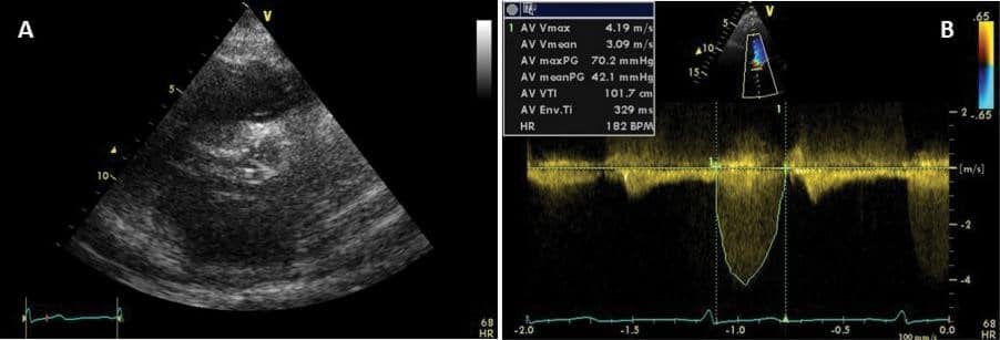
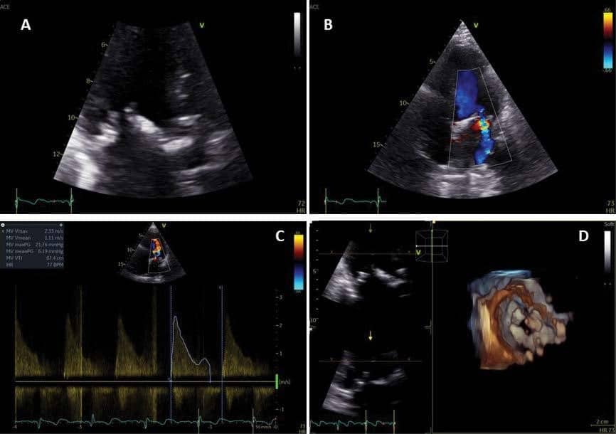
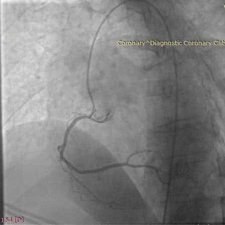
Transthoracic echocardiography quantified a progression of the aortic valve disease, now with severe stenosis and moderate regurgitation (Figure 1), mitral valve disease (mild stenosis and moderate regurgitation) (Figure 2), moderate tricuspid regurgitation and secondary pulmonary hypertension (PH) (estimated pulmonary artery systolic pressure (PAP) 91 mmHg), with normal left ventricular systolic function. In the absence of a clear etiology to explain the PH, right heart catheterization was performed and confirmed the PH, with a mean PAP of 50 mmHg and a PCW of 20 mmHg, confirming postcapillary PH. Therefore the patient does not meet the criteria for specific PAH treatment. Coronary angiography showed right-dominant circulation and an ostial stenosis of 60% of the right coronary artery (Figure 3).
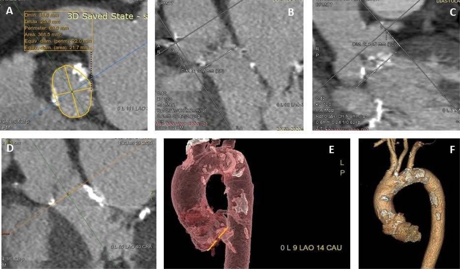
The patient has an indication for aortic valve replacement; in the context of past mediastinal radiotherapy the preferred method is transcatheter aortic valve implantation (TAVI). A cardiac computed tomography (CT), with systolic and diastolic multiphase recon-struction, was performed for better characterization of the aortic valve and annulus assessment, as well as for planning, landing zone calcification review and aortic root measurement, in order to perform TAVI procedure. The CT (Figure 4) emphasized the severe annular calcification and described a band of calcification in the posterior wall of the LV outfl ow tract extended from the left coronary cusp to the mitro-aortic junction (Figure 4D). Also, calcified plaques were described from the aortic root to the level of the first third of the descending aorta, suggestive for porcelain aorta (Figure 4F).
DISCUSSION
The present case is an illustration of the echocardiographic findings of radiotherapy related valvular disease, with fibrosis and calcification of the aortic root and aortic valve cusps, mitral valve annulus and the base and mid portions of the mitral valve leaflets, but with sparing the mitral valve tips and commissures (helping the differential diagnosis with rheumatic disease). The ostial lesions of coronary arteries are frequent after radiotherapy for HL, the most exposed being the left main stem and ostial segments of the circumflex and right coronary arteries. Survivors of HL have four- to seven-fold increased risk of coronary artery disease compared with the general population. Guidelines re-commend percutaneous coronary intervention only for ostial lesions of more than 70% before TAVI as a IIa indication, level of evidence C12. Porcelain aorta is frequently associated in this setting. Due to the extensive calcifications of the ascending aorta, mediastinal fibrosis, impaired wound healing and associated coronary artery disease, conventional cardiovascular surgery is frequently prohibited and, when having severe aortic stenosis, patients who underwent mediastinal radiotherapy are eligible for TAVI (class I indication, level of evidence B)12. Of note, PH is an independent risk factor for survival after TAVI. Nevertheless, TAVI was proven to lead to an acute improvement of nearly all invasively assessed variables in patients with PH, with a similar improvement in functional NYHA class compared to patients without PH, indicating a similar benefit among survivors13.
Conflict of interest: none declared.

Figure 1. Transthoracic echocardiography. A. Parasternal short axis view shows tricuspid aortic valve with limited systolic opening. B. Doppler interrogation of the aortic valve flow shows high peak velocity (4.2 m/s) and mean transaortic gradient (42 mmHg).

Figure 2. Transthoracic echocardiography. A. Zoomed image of apical long axis view showing mitral and aortic valve calcifications. B. Moderate mitral regurgitation. C. Color Doppler interrogation of the mitral valve shows elevated mean transmitral gradient (6.3 mmHg). D. 3D ventricular view of the mitral valve showing the limited maximal diastolic opening.

Figure 3. Coronary angiography showing 60% ostial stenosis of the right coronary artery.

Figure 4. Computer tomography (CT) planning of the TAVI procedure. A. Systolic measurements of the longest and shortest annulus diameters, area and perimeter. Severe annulus calcification with nodules protruding into annular lumen can be seen. B and C. Right and left coronary ostium height measurements, which in this case, are permissive for TAVI procedure. D. Subannular calcification extending from the left coronary cusp into the LVOT up to mitro-aortic junction. E. Fluoroscopic planning- provision of optimal fluoroscopic projection angulations based on CT. F. Porcelain aorta.
References
Jaworski C, Mariani JA, Wheeler G, Kaye DM. Cardiac complications of thoracic irradiation. J Am Coll Cardiol 2013;61:2319–2328.
2. Zamorano J, Lancellotti P, Muñoz D. 2016 ESC Position Paper on cancer treatments and cardiovascular toxicity developed under the auspices of the ESC Committee for Practice Guidelines: The Task Force for cancer treatments and cardiovascular toxicity of the Euro-pean Society of Cardiology (ESC). European Heart Journal, Volume 37, Issue 36, 21 September 2016, Pages 2768–2801.
3. Aleman BM, van den Belt-Dusebout AW, De Bruin ML, van‘t Veer MB, Baaijens MH, de Boer JP, Hart AA, Klokman WJ, Kuenen MA, Ouwens GM, Bartelink H, van Leeuwen FE. Blood. 2007 Mar 1;109(5):1878-86
4. Armstrong GT, Joshi VM, Ness KK, Marwick TH, Zhang N, Srivas-tava D, Griffin BP, Grimm RA, Thomas J, Phelan D, Collier P, Krull KR, Mulrooney DA, Green DM, Hudson MM, Robison LL, Plana JC. Comprehensive Echocardiographic Detection of Treatment-Related Cardiac Dysfunction in Adult Survivors of Childhood Cancer: Re-sults From the St. Jude Lifetime Cohort Study. J Am Coll Cardi-ol. 2015 Jun 16;65(23):2511-22.
5. Darby SC, Ewertz M, McGale P, Bennet AM, Blom-Goldman U, Bronnum D, Correa C, Cutter D, Gagliardi G, Gigante B, Jensen MB, Nisbet A, Peto R, Rahimi K, Taylor C, Hall P. Risk of ischemic heart disease in women after radiotherapy for breast cancer. N Engl J Med 2013;368:987–998.
6. Storey MR, Munden R, Strom EA, McNeese MD, Buchholz TA. Coronary artery dosimetry in intact left breast irradiation. Cancer J 2001;7:492–497
7. Hull MC, Morris CG, Pepine CJ, Mendenhall NP. Valvular dysfunc-tion and carotid, subclavian, and coronary artery disease in survi-vors of Hodgkin lymphoma treated with radiation therapy. JAMA 2003;290:2831–2837
8. Malanca M, Cimadevilla C, Brochet E, Iung B, Vahanian A, Messika-Zeitoun D. Radiotherapy-induced mitral stenosis: a three-dimen-sional perspective. J Am Soc Echocardiogr 2010;23:108 e101–102
9. Cutter DJ, Schaapveld M, Darby SC, Hauptmann M, van Nimwegen FA, Krol AD, Janus CP, van Leeuwen FE, Aleman BM. Risk of valvular heart disease after treatment for Hodgkin lymphoma. J Natl Cancer Inst 2015;107:djv008
10. Glanzmann C, Huguenin P, Lutolf UM, Maire R, Jenni R, Gumppen-berg V. Cardiac lesions after mediastinal irradiation for Hodgkin’s disease. Radiother Oncol 1994; 30:43–54
11. Hering D, Faber L, Horstkotte D. Echocardiographic features of ra-diation-associated valvular disease. Am J Cardiol 2003;92:226–230
12. Baumgartner H, Falk V, Bax JJ et al. on behalf of the Task Force for the Management of Valvular Heart Disease ofthe European Society of Cardiology (ESC) and the EuropeanAssociation for Cardio-Thoracic Surgery (EACTS). 2017 ESC/EACTS Guidelines for themanage-ment of valvular heart disease. Eur Heart J 2017; 38, 2739–2791
13. Schewel D, Schewel J, Martin J et al. Impact of transcatheter aortic valve implantation (TAVI) on pulmonary hyper-tension and clinical outcome in patients with severe aortic valvular stenosis. Clin Res Cardiol 2015; 104: 164–174.
 This work is licensed under a