A. Negoiţă, L. Zarma, D. Deleanu
„Prof. Dr. C.C. Iliescu” Emergency Institute for Cardiovascular Diseases, 258, Fundeni Street, Bucharest
Dr. Adrian Negoiță, „Prof. Dr. C.C. Iliescu” Emergency Institute for Cardiovascular Diseases, 258, Fundeni Street, Bucharest
We present the case of a 74 year old patient, hypertensive, dyslipidemic, with a personal history of angina going back several years, with an acute coronary syndrome without ST elevation which occurred in the past year, complicated with residual angina and heart failure symptoms. She is admitted accusing chest pain at rest, ceasing spontaneously after 10 minutes and dyspnea occurring during medium intensity efforts.
On admission the patient is in a good clinical condition, blood pressure (BP) 120/60 mmHg, pulse – 50 b/min. The EKG reveals sinus bradycardia, pulse – 45b/min, QS in V1 and V2 and an elevated, positive and symmetric T wave in V2 – V3;
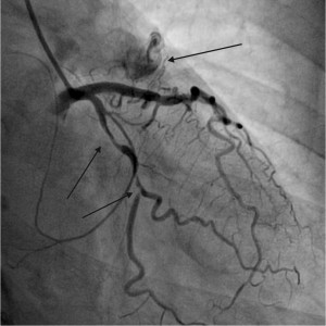
Figure 1. 80% stenosis of the circumflex artery (CX) in the first segment and 90% stenosis in the second and third segments of the circumflex artery (continuous arrow). The pulmonary artery accumulates contrast agent from branches emerging from the descending artery (dotted arrow).
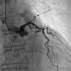
Figure 2. Contrast agent reflux in the aorta (continuous arrow), descending artery – pulmonary artery fistula (dotted arrow).
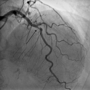
Figure 3. 80% stenosis of the anterior descending artery in the IInd segment immediately below the origin of the diagonal artery I.
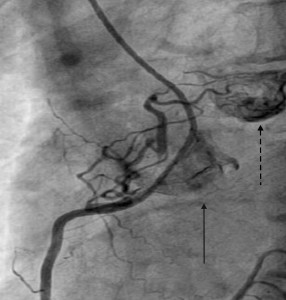
Figure 4. Contrast injection in the right coronary artery – contrast reflux in the aorta (continuous arrow), contrast captation – pulmonary artery via branches emerging from the proximal segment of the right coronary (dotted arrow).
The echocardiography finds normal dimensions for the LV, with a mild systolic dysfunction (Left Ventricle Ejection Fraction (LVEF) = 45-50%) and kinetic segment disorders in the antero-lateral and posterior walls.
The coronarography shows an 80% stenosis of the left anterior descending artery (LAD) II, immediately below the origin of the diagonal artery (DG) I, a 70-80%
stenosis of the circumflex artery (CX) I and 90% for CX II and III, right coronary artery without lesions. After contrast administration in the left coronary artery, a fistula between the proximal segment of the descending artery and the pulmonary artery is revealed. The same aspect is also present in the right coronary artery.
Conflict of interests: none.
 This work is licensed under a