Download PDF
https://doi.org/10.47803/rjc.2020.30.1.67
Mihai Roca1,2, Magda Mitu2, Radu-Sebastian Gavril1,2, Maria-Magdalena Leon Constantin1,2, Iulia-Cristina Roca1,3, Florin Mitu1,2
1 Grigore T. Popa” University of Medicine and Pharmacy, Iasi, Romania
2 Clinical Rehabilitation Hospital, Iasi, Romania
3 „Sf. Spiridon” Emergency Clinical Hospital, Iasi, Romania
Abstract: Cardiovascular rehabilitation represents a very important measure in post myocardial infarction patients for both, improving their quality of life and preventing other acute cardiovascular events. It is important to accurately assess functional capacity of patients after acute coronary events, in order to optimize the results of cardiac rehabilitation program. Cardiopulmonary exercise testing (CPET) represents the gold standard in functional capacity assessment. We present 3 cli-nical cases of post STEMI patients, with coronary revascularization interventions, addressed to cardiovascular rehabilitation. They underwent CPET evaluation at baseline and during rehabilitation program. This method proved important utility for individualization of cardiovascular rehabilitation program, as well as for monitoring the long term evolution after myocardial infarction.
Keywords: cardiopulmonary exercise testing, myocardial infarction, cardiac rehabilitation.
Rezumat: Recuperarea cardiovasculară reprezintă o măsură importantă la pacienţii post-infarct miocardic acut, atât din punct de vedere al efectelor de îmbunătăţire a calităţii vieţii, cât şi pentru prevenţia apariţiei altor evenimente cardiovas-culare. Pentru optimizarea rezultatelor programului de recuperare cardiovasculară este importantă evaluarea riguroasă a capacităţii funcţionale a pacienţilor după evenimentul coronarian acut. Testarea cardiopulmonară de efort (CPET) reprezintă standardul de aur în evaluarea capacităţii funcţionale. Prezentăm trei cazuri clinice ale unor pacienţi cu STEMI şi intervenţii de revascularizare coronariană, la care s-a indicat recuperarea cardiovasculară. La aceşti pacienţi s-a realizat evaluarea prin CPET atât la iniţiere cât şi pe parcursul programului de reabilitare cardiovasculară. Metoda s-a dovedit utilă pentru indivi-dualizarea programului de recuperare cardiovasculară dar şi pentru monitorizarea evoluţiei pe termen lung la pacienţii cu infarct miocardic.
Cuvinte cheie: testare cardiopulmonară de efort, infarct miocardic, recuperare cardiovasculară.
INTRODUCTION
Although myocardial infarction remains a common event in patients with cumulative cardiovascular risk factors, the increasing accessibility to modern the-rapeutic resources of myocardial revascularization, determined a signifi cant improvement of surviving1. However, patients surviving an acute coronary event need to be included in a cardiovascular rehabilitation (CR) program, to restore their functional capacity and quality of life, to control risk factors and to prevent the recurrence of acute cardiovascular events2.
It is important to individualize the parameters of aerobic exercise training within CR program, conside-ring clinical and functional particularities of the patient.
Currently, the gold standard in functional capa-city assessment is cardiopulmonary exercise testing (CPET), recommended by clinical guidelines for CR, in order to maximize benefits and to minimize risks associated to aerobic exercise training3-7. In addition, CPET represents the most rigorous assessment of the effectiveness of CR program8,9.
In this paper we present three clinical cases to exemplify CPET using in CR programs, highlighting the benefits obtained, according to the clinical particulari-ties of each case.
CASE SERIES
Case 1
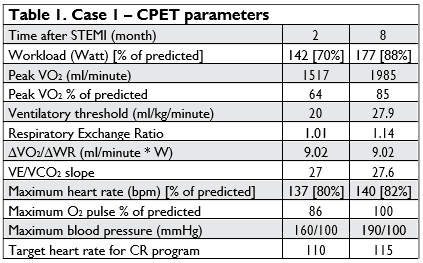
A 48-year-old male was known for ten years with es-sential hypertension, type 2 diabetes mellitus under oral therapy and mixed dyslipidemia. The patient was admitted to Clinical Recovery Hospital Iasi, for phase II CR, two month after an anterior STEMI, treated by percutaneous coronary intervention (PCI), with drug eluting stent implantation on the left anterior descen-ding artery. At the admission, the patient presented slight limitation during ordinary activity. Echocardio-graphy revealed a left ventricular apical hypokinesia and a left ventricle ejection fraction limited to 50%.
Functional capacity was assessed by CPET, before initiating CR. The most important CPET parameters were: maximum oxygen consumption (peak VO2), maximum workload (WR [Watt]), ventilatory (ana-erobic) threshold (AT), oxygen (O2) pulse, relation between VO2 and workload (∆VO2/∆WR slope), re-lation between minute ventilation (VE) and carbon di-oxide production (VE/VCO2 slope), maximum heart rate, maximum blood pressure (Figure 1, 2, 3, 4).
CPET revealed a pattern of cardiac moderate func-tional limitation (peak VO2 64% of predicted value) and determined a target heart rate of 110 beats per minute (bpm), for aerobic exercise training during CR program. The main results are presented in Table 1 (two month after the STEMI). The patient continued a long term ambulatory CR program. After 6 month, CPET assessment revealed signifi cant improvement of the functional capacity (Table 1 – 8 month after the STEMI).


Figure 1. Peak VO2 detection (case 1): maximum oxygen consumption recorded during progressive intensity exercise; ventilatory (anaerobic) threshold (AT) detection: During progressive increasing of exercise intensity, at a given workload, oxygen supply to the muscle does not meet the oxygen require-ments. This imbalance determines the necessity of anaerobic glycolysis in order to provide muscle energy, resulting in lactic acid production. Conversion of lactic acid to lactate, results in an excess of CO2 production, which is revealed on graphical representation of trend for VO2 and VCO2 (anaerobic threshold).

Figure 2. Ventilatory threshold detection applying ventilatory equivalents method (case 1): At the anaerobic threshold, a physiological increase in ventilation (VE) simultaneously appears, to eliminate the excess CO2 (ventilatory threshold). It is the point at which, an increase of the ventilatory equivalent for oxygen (VE/VO2) occurs, without an increase of ventilatory equivalent for carbon dioxide (VE/VCO2).

Figure 3. Target heart rate detection for aerobic training within cardiovascular rehabilitation (case 1): Ventilatory threshold marks the upper limit of light to moderate – intensity effort domain. Light to moderate-intensity training is the most indicated for cardiovascular rehabilitation of cardiac patients with a markedly reduced exercise capacity, for those with high exercise-related risk, or recent hemodynamic decompensation, including patients after myocardial infarction3. The heart rate corresponding to ventilatory threshold represents the target heart rate during aerobic exercise training: 115 bpm for case 1.

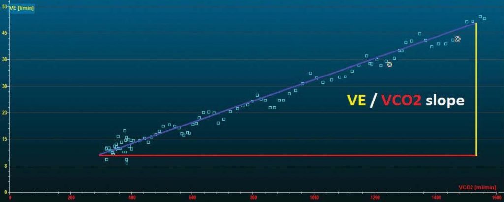
Figure 4. VE/VCO2 slope calculation (case 1): This parameter represents an index of ventilator efficiency, with a normal threshold <30, which can be ex-ceeded in heart failure or pulmonary hypertension. This parameter is not influenced by age or sex and presents high test-retest reliability, being uninfluenced by exercise testing protocol 4. For case 1, VE/VCO2 slope presented a normal value of 27.

Case 2
A 35-year-old male, smoker, overweight, known with dyslipidemia, was admitted to our clinic, for phase II CR, one month after an anterior STEMI, treated by PCI. Echocardiographic examination at hospital admis-sion revealed a left ventricular apical hypokinesia and a left ventricle ejection fraction of 50-60%.
The results of CPET were consistent with a car-diac pattern of moderate-severe functional limitation: decreased peak VO2 and ventilatory threshold; decre-ased maximum O2 pulse; normal ventilatory reserve (Table 2 – one month after the STEMI).
The patient initiated CR in our clinic, continuing an ambulatory CR program. Two month later, a second CPET revealed slight improvement of the most im-portant parameters (Table 2 – CPET parameters at 3 month after the STEMI). A third CPET assessment was done four month later, proving signifi cant impro-vements of workload, maximum oxygen consumption and maximum heart rate. However, the results reve-aled a significant negative trend, apparently paradoxi-cal, for two key parameters: ventilatory threshold and maximum oxygen pulse (Table 2 – CPET parameters at 7 month after the STEMI). The next CPET assess-ment, done at 15 month, revealed important changes in functional capacity. Although work rate level seems to improve, peak VO2 had a slight decrease, while ven-tilatory threshold and maximum heart rate presented a signifi cant negative trend (Table 2 – CPET parame-ters at 22 month after the STEMI).
Inline with CPET changes, echocardiography reve-aled left ventricular apical hypokinesia, involving in-terventricular septum and left ventricle lateral wall, suggesting left ventricle apical aneurysm. Furthermo-re, left ventricle ejection fraction decreased to 40-45%, compared to previous echographic exam.
Case 3
A 56-year-old male, smoker, was known with chronic coronary artery disease (left anterior descending ar-tery and left main trunk lesions), hypertension, mixed dyslipidemia, grade I obesity. The patient was admitted in our clinic, to be included in phase II CR program, five month after anteroseptal STEMI and coronary artery bypass grafting (CABG). At the admission, the patient presented a moderate limitation during ordi-nary activity. Echocardiographic examination revealed left ventricular regional wall motion abnormality with ejection fraction of 50%.
Maximum oxygen consumption, and ventilatory threshold assessed at CPET, corresponded to a mo-derate-severe alteration of functional capacity, while O2 pulse was 76% of predicted maximum (Table 3 – 5 month after the STEMI). The results of CPET presen-ted above were consistent with a cardiac pattern of moderate-severe functional limitation.
The patient initiated CR in our clinic, continuing an ambulatory CR program. One year later, the results of CPET proved significant improvements of functio-nal capacity, from moderate-severe alteration to mild-moderate alteration (Table 3 – CPET parameters at 17 month after the STEMI). Echocardiography reve-aled left ventricular segmental hypokinesia, with pre-served ejection fraction.

DISCUSSION
We presented 3 clinical cases of post STEMI patients addressed to CR. The fi rst two cases were young ma-les, successfully treated by PCI.
For all three cases, CR program was initiated with 20 minutes sessions of mild, progressively increased to moderate intensity, continuous aerobic exercise training, aiming the target heart rate individually deter-mined by CPET. This test allowed the optimal setting of intensity for exercise training. Training session was preceded and followed by warm-up and cooling down 10 minutes periods, respectively.
First case presented higher baseline aerobic capacity and O2 pulse, and a very good long term improvement of these parameters during CR program. However, the second case presented signifi cant reductions of baseline levels for aerobic capacity and O2 pulse, and a failure in improvement of these parameters during long term evolution, despite carrying out CR program.
This lack of efficiency of CR in second case was concordant with echocardiographic aspects of unfavo-rable myocardial remodeling with left ventricle apical aneurysm and ejection fraction decreasing. However, alterations of maximum O2 pulse during CPET, prece-ded echographic changes. Our results are inline with those of some clinical studies which proved that selec-ted CPET parameters (as peak VO2, ventilatory thre-shold, O2 pulse) seem to be highly sensitive to changes in cardiac function following PCI, significantly better than conventional stress ECG10,11.
In the second clinical case, ventilatory threshold was the best predictor of unfavorable evolution, pre-senting the earliest and the most significant negative trend, which preceded echocardiographic alterations.
The third clinical case represents a patient addressed to CR post STEMI and CABG. Unlike second clinical case, the long term evolution was favorable, with sig-nifi cant functional improvement during CR, despite baseline low values of peak VO2, ventilatory threshold and O2 pulse. These parameters presented impor-tant CR related improvement. However, there is a lack of clinical researches referring the predictors of long term cardiac changes in patients with STEMI and CABG following CR12.
Considering the results previously obtained in other clinical studies, a lower baseline aerobic capacity and a more reduced baseline ejection fraction, represent predictors of a higher functional capacity improvement after exercise based CR, among myocardial infarction survivals, irrespective of the management modality for acute coronary event13,14.
CONCLUSIONS
CPET greatly enhance the evaluation of patients addressed to cardiovascular rehabilitation. This test is essential for optimizing the parameters of aerobic exercise training, in order to maximize benefits and to minimize the risk for CR program. Furthermore, CPET repeatedly done, during and after CR program, facilitates the objectification of functional cardiovas-cular benefits, within this program, and allows a long term prognostic assessment. Prognostic value of CPET may exceed the evaluation methods in resting condi-tions, such as cardiac echography and ECG. However, the role of CPET in CR is not completely defined, new clinical studies being necessary.
Conflict of interest: none declared.
References
1. Neumann FJ, Sousa-Uva M, Ahlsson A, Alfonso F, Banning AP, Bene-detto U, Byrne RA, Collet JP, Falk V, Head SJ, Jüni P, Kastrati A, Koller A, Kristensen SD, Niebauer J, Richter DJ, Seferovic PM, Sib-bing D, Stefanini GG, Windecker S, Yadav R, Zembala MO; ESC Sci-entific Document Group. 2018 ESC/EACTS Guidelines on myocar-dial revascularization. Eur Heart J 2019;40(2):87-165.
2. Corrà U, Mendes M, Piepoli M, Saner H. Future perspectives in car-diac rehabilitation: a new European Association for Cardiovascular Prevention and Rehabilitation Position Paper on ‘secondary preven-tion through cardiac rehabilitation’. Eur J Cardiovasc Prev Rehabil 2007;14(6):723-5.
3. Varga A, Tilea I, Palermo P. Cardiopulmonary exercise test in clini-cal practice, evaluation of myocardial ischemia. Romanian Journal of Cardiology 2019;29(3):390-398.
4. Caloian B, Pop D, Guşetu G, Zdrenghea D. The role of cardiopul-monary exercise testing in the initial evaluation of patients wearing intracardiac devices submitted to cardiac rehabilitation. Balneo Re-search Journal 2017;8(4):206-211.
5. Lollgen H, Leyk D. Exercise testing in sports medicine. Dtsch Arz-tebl Int 2018;115:409-16.
6. Mezzani A. Cardiopulmonary exercise testing: basics of methodol-ogy and measurements. Ann Am Thorac Soc 2017;14:S3-11.
7. Gherasim D. Rolul testului de efort în programele de recuperare. În: Recuperare şi prevenţie cardiovasculară. Editor: Zdrenghea D. Edi-tura Clusium, Cluj-Napoca, 2008, 64-92.
8. Mezzani A, Hamm LF, Jones AM, McBride PE, Moholdt T, Stone JA, Urhausen A, Williams MA. Aerobic exercise intensity assessment and prescription in cardiac rehabilitation: a joint position statement of the European Association for Cardiovascular Prevention and Re-habilitation, the American Association of Cardiovascular and Pulmo-nary Rehabilitation and the Canadian Association of Cardiac Reha-bilitation. Eur J Prev Cardiol 2013;20(3):442-67.
9. Balady GJ, Arena R, Sietsema K, Myers J, Coke L, Fletcher GF, For-man D, Franklin B, Guazzi M, Gulati M, Keteyian SJ, Lavie CJ, Macko R, Mancini D, Milani RV. Clinician’s Guide to cardiopulmonary exer-cise testing in adults: a scientific statement from the American Heart Association. Circulation 2010;122(2):191–225.
10. Inbar O, Yamin C, Bar-On I, Nice S, David D. Effects of percutane-ous transluminal coronary angioplasty on cardiopulmonary respons-es during exercise. J Sports Med Phys Fitness 2008;48(2):235-45.
11. Chaudhry S, Arena R, Bhatt DL, Verma S, Kumar N. A practical clinical approach to utilize cardiopulmonary exercise testing in the evaluation and management of coronary artery disease: a primer for cardiologists. Curr Opin Cardiol 2018; 33(2): 168–177.
12. Suzuki Y, Ito K, Yamamoto K, Fukui N, Yanagi H, Kitagaki K, Konishi H, Arakawa T, Nakanishi M, Goto Y. Predictors of improvements in exercise capacity during cardiac rehabilitation in the recovery phase after coronary artery bypass graft surgery versus acute myocardial infarction. Heart Vessels 2018;33(4):358-366.
13. Aguiar Rosa S, Abreu A, Marques Soares R, Rio P, Filipe C, Rodrigues I, Monteiro A, Soares C, Ferreira V, Silva S, Alves S, Cruz Ferreira R. Cardiac rehabilitation after acute coronary syndrome: Do all pa-tients derive the same benefit? Rev Port Cardiol 2017;36(3):169-176.
14. Vilela EM, Ladeiras-Lopes R, Ruivo C, Torres S, Braga J, Fonseca M, Ribeiro J, Primo J, Fontes-Carvalho R, Campos L, Miranda F, Nunes JPL, Gama V, Teixeira M, Braga P. Different outcomes of a cardiac rehabilitation programme in functional parameters among myocar-dial infarction survivors according to ejection fraction. Neth Heart J 2019;27(7-8):347-353.
 This work is licensed under a