Elena Parvu1, Stefan Ailoaie2, Daniel Gafitescu2, Liviu Marian Stoica2, Gabriela Creanga2, Mihaela Grecu2
1 Johnson and Johnson Company
2 „Prof. Dr. George I.M. Georgescu” Institute for Cardiovascular Diseases, Iasi, Romania
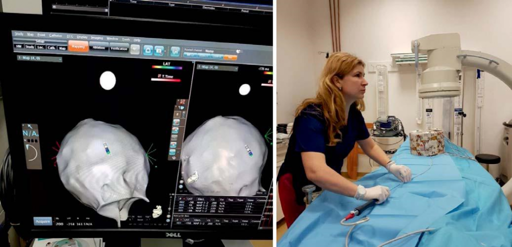
A piggy bank was reconstructed as right atrium of an imaginary patient, during a training process using 3D Carto Mapping System, Biosense Webster Company.
The following story took place in Cardiovascular Diseases Institute “Prof. Dr. George I.M. Georgescu” Iasi, an EHRA recognized training center for young electrophysiologists, under the coordination of Mihaela Grecu – electrophysiologist and trainer within the institution.
After a usual 3D ablation was performed, the ma-pping catheter used during the procedure was not dis-carded, but properly washed and used to create a fast-anatomical map outside the patient. The main goal was to train healthcare professionals (young EP, resident physicians, students etc.) who want to understand 3D mapping concepts in a practical manner, at a very basic level in a low-cost manner.
The team consisting of 1 CARTO technician, 2 principal medical nurses and 2 physicians in training resi-dents simulated a patient using the minimum require-ments for such an activity and reusing materials from the previous ablation procedure. The patient was simulated as a fixed cardboard box, the patient’s heart was a piggy bank with a basal and one upper opening and the inferior vena cava was simulated by the protective sleeve from the original catheter’s packaging.
The box was placed on the patient table, on the approximate position of the patient’s chest, within the location pad’s perimeter; the piggy bank (heart) was placed and secured with adhesive tape inside of the box and the protective tube was attached to its lower opening. The two sets of patches were properly pla-ced under and above the box. A metallic wire was added to make a connection between the patches, creating this way the electrical contact between them.
The mapping catheter (8F, irrigated Smarttouch Thermocool) was then connected to the PIU and the CARTO 3 system did not display any errors, recogni-zing the “patient” as a real one. After having the catheter advanced into the tube (inferior cave vein) to the piggy bank (heart), the 3D anatomical construction was started. In this way, the EP trainee could practice his catheter maneuverability (rotation, curves etc) and get use to the 3D projections and orientation.
As a final challenge, to test the understanding af-ter the anatomical construction, the mapping catheter had to be inserted into the upper opening of the piggy bank – about 7 mm in width and 30 mm in length. The test was passed with success!
An enthusiastic equip can find ingenious and very low-cost approach to practice 3D mapping catheter maneuverability outside the patient.
DISCUSSION
This method of patient simulation for the CARTO 3D system can be used to prepare young electrophysiologists, to learn handling, the positioning and flipping – deflection of the catheters. It is a simple and inexpensive method to understand catheter movement in 3D space and to practice the manual force that is needed to rotate and move the catheter into a small anatomi-cal space.
The simulation system presented does not replace a high-fidelity hybrid simulator (Procedicus VIST, ver-sion 7.0, Mentice AB Gothenburg, Sweden for Biosen-se Webster) used in specialized training centers1.
Conflict of interest: none declared.
References
1. Roberto De Ponti, Raffaella Marazzi, Lorenzo A. Doni, Claudio Tam-borini, Sergio Ghiringhelli, Jorge A. Salerno-Uriarte: Simulator train-ing reduces radiation exposure and improves trainees’ performance in placing electrophysiologic catheters during patient-based proce-dures. Heart Rhythm, Volume 9, Issue 8, August 2012, Pages 1280-1285.
 This work is licensed under a