A. Negoiţă, D. Deleanu
Article received on the 7th of August 2012. Article accepted on the 27th of August 2012.
„Prof. Dr. C.C. Iliescu” Emergency Institute for Cardiovascular Diseases, 258 Fundeni Street, Bucharest
„Prof. Dr. C.C. Iliescu” Emergency Institute for Cardiovascular Diseases, 258 Fundeni Street, Bucharest
We present the case of a 73 year old hypertensive and dyslipidemic patient, under immunosuppressive treatment for rheumatoid polyarthritis, who presented 7 hours after onset of typical angina.
Upon admission the patient is hemodynamic stable, BP = 160/90 mmHg, heart rate = 75 bpm, atrial fibrillation is diagnosed based on the electrocardiogram, V3 – V6 ST segment elevation, DI, aVL of maximum 2 mm, q waves present in DI and aVL and echocardiographic wall motion disorders are highlighted at the apex and apical segments of the left ventricular wall.
The coronarography indicates an acute thrombotic occlusion of a branch from the second diagonal artery, possibly embolic, with TIMI flux 0 (Figure 1), without any other lesions on the other blood vessels. It is decided to perform the angioplasty at this level, without succeeding in passing with the thrombus aspiration catheter through the occlusion zone. The median portion of the vessel is pre-dilated with a 2.0/20 mm balloon (Figure 2). Distal embolisation can be observed at the control injection of contrast. The distal occlusion zone is pre-dilated with the same balloon and later, an extravasation of contrast substance can be observed in the distal segment of the vessel, with a particular, possibly subepicardial aspect (Figure 3). Immediately the same balloon is inflated on the perforation site for 15 minutes, with the remission of the effraction zone (Figure 4).
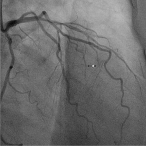
Figure 1. Acute thrombotic lesion of a branch of the IInd diagonal artery (arrow).
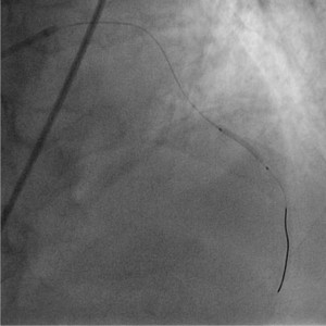
Figure 2. Crossing the occlusion area with a 0.014 microscope and pre-dilation with a 2.0/20 mm balloon.
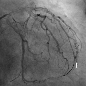
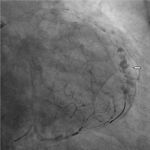
Figure 3. A) we can see the area where the contrast substance extravasation, distally on the branch of the second diagonal artery. B) particular aspect of the extravessel aspect of contrast substance, possibly located subepicardial.
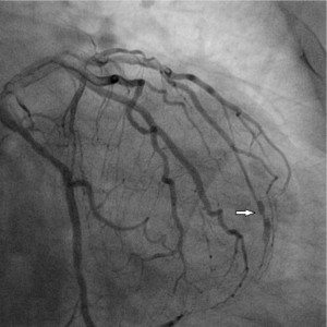
Figure 4. Final results, remission of the effraction zone, with an aneurismal aspect at this level (the arrow) and good distal flux.
The patient’s evolution was favorable during hospitalization, lacking any recurrence of the anginous episode, hemodynamically stable, without visible pericardic fluid at the ecocardiographic evaluation and with an amelioration of the segmentary kinetics disturbances.
Conflict of interest: none declared.
 This work is licensed under a