Andreea Varga1, Claudia Floriana Suciu1, Dragos-Gabriel Iancu2, Dorina Nastasia Petra1, Ioan Tilea1,2
1 University of Medicine, Pharmacy, Sciences and Technology, Targu Mures, Romania
2 Department of Cardiology, Emergency Clinical County Hospital, Targu Mures, Romania
Abstract: Background – Systemic lupus erythematosus (SLE) affects young women, in smaller degree young men. Particularly particular male SLE phenotype presents poor prognosis and miscellaneous organ damage. Subsequent SLE atherosclerosis along with unremarkable traditional atherogenic risk factors determines severe coronary artery disease in long-standing SLE disease. The clinical profile, diagnostic and management algorithm are considered, as well as a brief review of current literature. Case presentation – A 66-year-old Caucasian male with SLE diagnosed in 1998 was admitted for extended cardiovascular assessment. Due to severe pancytopenia related to SLE specific therapy, the patient was placed on a time-adjusted dosage of corticosteroids. Thorough the time, patient developed dilated cardiomyopathy, atrial fi brillation (AF) and progressive heart failure (HF), in the absence of a close cardiac follow-up. Cardiac catheterization revealed severe coronary artery disease (CAD) without evidence of marked atherogenic risk factors. However, the patient experienced non-traditional atherosclerotic risk factors such as hyperhomocysteinemia (HH), increased oxidized low-density lipopro-tein cholesterol (OxLDL), and hyperphosphatemia. Conclusion – Cardiac involvement is a frequent manifestation of SLE, associated with a high morbimortality. Management of CAD, AF and HF with mild reduced ejection fraction (HFmrEF) in a SLE patient with severe chronic kidney disease (CKD) is challenging. An individualized strategy of close follow-up in specific autoimmune disease patients is needed.
Keywords: systemic lupus erythematosus, coronary artery disease, heart failure, hyperhomocysteinemia, oxidized low-density lipoprotein cholesterol.
INTRODUCTION
Systemic lupus erythematosus (SLE) is a chronic au-toimmune inflammatory disorder that mainly affects young women, and to a smaller degree, young men as well. In elderly SLE patients with long-standing di-sease, coronary artery disease (CAD) was confirmed as a common cause of mortality1,2. Male SLE patients display particular features, have a worse prognosis and multiple organ damage3. Furthermore, SLE patients have an up-to 3.39 increased risk for cardiovascular disease (CVD) compared to non-SLE individuals, par-ticularly young women4,5.
SLE risk factors for CVD, specifi cally secondary to atherosclerosis surpass the traditional atherosclerotic risk factors. Dyslipidaemia is commonly linked to cor-ticosteroid treatment1.
Specific SLE dysregulated immune responses are associated with both increased risk of premature atherosclerotic plaques development along with an increased risk of plaque rupture. Consequently, lu-minal thrombosis, plaque erosions, and calcified no-dules have a high risk of occurrence, thus triggering acute coronary syndrome (ACS). Coronary arteritis and thrombosis related to coronary aneurysms or an-tiphospholipid syndrome can also lead to ACS6.
Kidneys are affected in about 50% SLE patients, with a higher risk of hyperhomocysteinemia and increased serum phosphate levels7. In particular, patients with lupus nephritis (LN) exhibit notable incremental CV mortality compared to non-LN SLE patients8
CASE PRESENTATION
A 66-year-old Caucasian male with long-standing SLE (diagnosed in 1998, met SLICC criteria in 2012) was admitted in an university-based hospital for extended cardiovascular assessment.
Lupus nephritis, CKD and secondary hypertension were diagnosed in 2004. A percutaneous renal biopsy was never performed; therefore, the classifi cation of LN accordingly to “The 2003 International Society of Nephrology/Renal Pathology Society classification of LN” was inapplicable9.
Severe pancytopenia related to administration of antimalarial agents (Hydroxychloroquine) or immuno-suppressive (Methotrexate or Azathioprine) was re-corded. Due to disease activity and despite emergent side-effects of long-term corticotherapy, Prednisone was prescribed, time-adjusted dosage, starting with 70 mg daily in 2004, down-titrated to 10 mg (from 2005). In February 2018 patient’s SLEDAI-2K (SLE Disease Activity Index) score was 32, as a consequence of sig-nificant organ involvement after a long evolution of the disease: visual disturbances, arthritis, urinary casts, hematuria, pyuria, rash, and alopecia (Figure 1).
Markers for secondary hyperparathyroidism and renal osteodystrophy were present: increased serum phosphate levels, elevated intact parathormone serum levels, and reduced 1.25-dihydroxy vitamin D.
With inconstant CV follow-up, in 2017 ischemic di-lated cardiomyopathy, mitral regurgitation (MR), AF, HFmrEF NYHA functional class III, and severe CKD were diagnosed.
Dyslipidaemia status was unremarkable throughout disease evolution (Figure 2).
On admission, physical examination revealed moon face, moderate lower limbs edema. An irregular car-diac rhythm (heart rate: 100 bpm, peripheral pulse: 80 bpm), mild MR murmur, left anterior tibial peripheral pulse absence were found. A diminished bilateral pedal pulse was present.
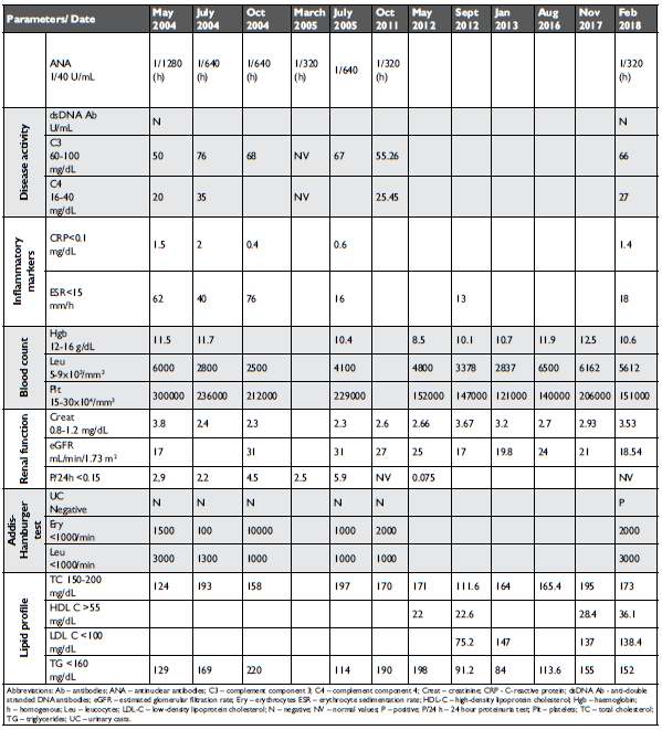
Laboratory tests showed mild anemia, NT-proBNP value of 32,604 pg/mL (normal values <210.0 pg/mL), positive ANA antibodies (1:320, normal range <1:40), a homogenous pattern and normal titres for anti-dsD-NA Ab, anti Smith Ab, anti SSA Ab, anti SSB Ab, anti RNP 70 Ab, anti ribosomal P protein Ab, anticardioli-pin Ab, anti phospholipid Ab (Ab against beta 2-glyco-protein I, cardiolipin, phosphatidylinositol, phospha-tidylserine, phosphatidic acid).
Complement C3 and C4 were within the normal range. Elevated serum C – reactive protein levels (1.4 mg/dL, normal values <0.33 mg/dL) with normal eryth-rocyte sedimentation rate were found. Severe CKD was confi rmed (eGFR: 17 mL/min/1.73m2), with a nor-mal 24-hour urine protein. Lipid profile, assayed using automated systems (Cobas, Roche Diagnostics) was not remarkable: normal values of total cholesterol and very low-density lipoprotein (VLDL) cholesterol, bor-derline triglycerides (190 mg/dL). Hyperhomocystei-nemia (21.95 μmol/L, normal values <10 μmol/L) and elevated OxLDL (94.6 U/L, normal range: 63.23 +/-16.23 U/L) were listed.
Laboratory test results throughout disease pro-gression are depicted in Table 1:
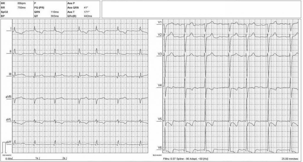
ECG recording identified AF with moderate ventricular response, LHV, myocardial ischemia patterns in anterolateral leads.
Echocardiographic data (Vivid E9™, General Elec-tric Company, Boston, MA, USA) described hyper-trophy (LHV) and dilation of the left ventricle with HFmrEF, moderate mitral and pulmonary regurgitati-on, and mild pulmonary systolic hypertension (Figures 4, 5, Table 2).
Lower limbs Duplex ultrasound scanning (HD 11XE™, Koninklijke Philips Electronics N.V., Nether-land) identified distal occlusion of the left anterior ti-bial artery; with no significant atherosclerotic plaques in other predictable arterial areas.
Unexpected severe CAD (triple vessel disease) was found on coronary angiogram (Allura Xper FD10 X-ray system, Koninklijke Philips Electronics N.V., Netherland): significant left main distal stenosis, sequen-tial stenosis and aneurysms of left anterior descending artery, occlusion of circumfl ex artery, significant ste-nosis of marginal branch and vertical segment of right coronary artery, with retrograde filling of left circum-flex artery (Figures 6, 7, 8).
Coronary CT angiogram or left ventriculography were not performed (severe CKD).
Either conservative approach or concomitant me-dical therapy and myocardial revascularization proce-dures (CABG vs. incomplete PCI) were considered, despite SYNTAX II score ≥3210. An unacceptable surgical risk based on EuroSCORE II and STS scores and concomitant administration of corticosteroids were assumed by a high-volume heart-team. Succes-sive percutaneous coronary interventions procedures were deemed as inappropriate. Finally, a conservative approach (Bisoprolol 5 mg bid, Amlodipine 10 mg od, Furosemide 40 mg bid and potassium supplementati-on) was offered in patient-centered therapeutic regi-men of HFmrEF, secondary hypertension, and CKD.
With no prior medical history of an acute left infe-rior limb ischemic episode, an embolic etiology of the left anterior tibial artery occlusion was ruled out.
Anticoagulation regimen the presence of AF EHRA II score and CHA2DS2-VASc=4, HAS-BLED=4 scores had to be decided. NOAC’s (Apixaban, Rivaroxaban, Dabigatran) were excluded with respect to concomi-tant severe CKD and prolonged corticosteroid treat-ment. Vitamin K antagonist (Acenocoumarol) was proposed as a life-long anticoagulation INR-adjusted regimen.
Therapeutic regimen for SLE consisting of Predni-sone 10 mg od along with Atorvastatin 80 mg od was recommended. SLE induced atherosclerosis and documented CAD required statins, benefits of this pharma-cotherapy being indisputable11,12.

Figure 1. SLE Disease Activity Index (SLEDAI-2K) between 2004-2018.

Figure 2. Lipid profile between 2004-2018.


Figure 3. ECG recording at admission.


Figure 4. Enlarged left ventricle (parasternal long-axis view).
Abbreviations: Ao – aorta, LA – left atrium, LV – left ventricle, RV – right ventricle.

Figure 5. Moderate mitral regurgitation (apical 4 chambers view).
Abbreviations: LA – left atrium, LV – left ventricle, RA – right atrium, RV – right ventricle, MR – mitral regurgitation.

Figure 6. Left coronary angiogram: LM – left main coronary artery, LAD
– left anterior descending artery. Arrows indicate LAD aneurysms (1) and severe stenosis (2).

Figure 7. Left coronary angiogram: LM – left main coronary artery, LAD – left anterior descending artery, Cx – circumflex artery. Arrows indicate LM (1) and marginal branch (2) stenosis, respectively Cx occlusion (3).

Figure 8. Right coronary angiogram: RCA – right coronary artery, Cx
– circumflex artery. Arrows indicate RCA stenosis (1) and collaterals to occluded Cx (2).
DISCUSSIONS
In the SLE population, traditional risk factors for CVD are reinforced by cardiovascular risk factors secon-dary to SLE such as the presence of antiphospholipid antibodies (APL) that play an essential role in throm-bogenesis and presence of antibodies against HDL cholesterol13.
At the time of diagnosis, 36.3% adult SLE patients experienced dyslipidemia, while 60% develop altered lipidic profiles after 3 years assessment14,15. Specific SLE dyslipidemic profi les can either be related to ac-tive disease16 or secondary to corticotherapy17,18. Ne-vertheless, a clear distinction between the two profi – les cannot be established.
Lupus patients, particularly those with active di-sease, present a specific dyslipidemic profile with low HDL cholesterol, high triglycerides, increased VLDL cholesterol, and normal to high LDL cholesterol19. Abnormal serum homocysteine20 and an important proinfl ammatory state also contribute to atheroscle-rotic disease in SLE patients as early atherosclerosis is acknowledged as the primary cause of mortality2.
In healthy individuals, hyperlipidemia is lowered by means of HDL and other newly discovered adaptive mechanisms such as an endogenous molecule, Del-1 (Developmental Endothelial Locus-1) that can bind to oxLDL and inhibit binding to oxLDL receptors21. The-se mechanisms are not suffi cient in SLE patients due to the presence of persistent dyslipidemia, renal di-sease, oxidative stress that leads increased production of OxLDL, and occurrence of antibodies against the protein contents of HDL that cancel the protective effects of HDL22.
Increased disease activity in SLE patients has been recognized as a potential non-traditional atheroscle-rotic risk factor, as the incidence of cardiovascular events was reported to be elevated in these indivi-duals23. However, due to important discrepancies between the studies design and the activity indices used, inconclusive results have been reported and an increased disease activity in SLE has not been validated yet as a potential cardiovascular risk factor24.
Our patient presented a constant active SLE with an increased SLEDAI score after a long disease evo-lution due to considerable organ damage and insuf-ficient immunosuppressive treatment. In accordance with data from previous studies25, his lipid profile was modified, most likely as a result of increased disease activity but also long-term corticotherapy and CKD; however, lipid values were not as excessive in order to justify the significant CAD. Important CAD burden requires secondary intensive statin treatment, ator-vastatin 80mg daily12. In the SATURN trial, high statin doses proved to have a signifi cant effect on plaque re-gression in patients with CAD. Due to his important plaque burden with increased risk of coronary plaque rupture, we considered that the patient would benefit most from the recommended doses of atorvastatin26.
Glucocorticoid treatment has been long known to correlate with CVD and atherosclerosis in the SLE population27. A 10 mg/day or more of corticostero-ids correlates with an incremental risk of CV events. Long-term use of 5 mg daily of Prednisone or less is associated with an acceptable low level of CV damage, while the use of more than 10 mg daily of Predniso-ne is associated with an increased level of CV impair-ment28.
Hyperhomocysteinemia (HH) represents an in-dependent risk factor for atherosclerosis leading to premature CVD29, as it can enhance atherosclerosis by increasing oxidative stress and maintaining a pro-infl ammatory state30. Common causes of HH are age, vitamin deficiency, impaired renal function with redu-ced glomerular filtration rate and mutations in genes responsible for the homocysteine metabolism. HH has been shown to correlate with premature atheroscle-rosis31, coronary artery calcification32,33 progression of atherosclerosis34,20, disease severity and thrombotic risk35, in SLE patients, with and without renal function impairment. Detected mild homocysteine increases undoubtedly contributed to his advanced CAD. Even though HH lowering therapy is available (folic acid and B-vitamins), the outcome on atherosclerosis progres-sion is discouraging. However, in placebo-controlled clinical trials, mild HH level are lowered, unimpressive results have been reported, as HH lowering therapy failed to have a significant impact on atherosclerotic CVD and/or athero-thrombotic CAD36.
Kidney disease is recognized as a consistent athe-rosclerotic risk factor in SLE patients, particularly in cases with poor control. Increased oxidative stress induced by LN and advanced CKD is attributable. In the general population, lower eGFR is associated with a higher CVD risk-related death37. In SLE population, in the first 5 years after diagnosis, approximately 60% will develop kidney disease38.
Kidney involvement was present in our patient at SLE diagnosis; however, the nature of the kidney disease could not be determined due to the lack of a kidney biopsy. Nevertheless, with longstanding kidney disease he developed renal osteodystrophy and secon-dary hyperparathyroidism. Elevated serum phosphate is known to be associated with increased arterial sti-ffness and coronary atherosclerotic plaque calcificati-on in patients with normal kidney function39. Higher phosphate serum levels may lead to the formation of calcified atherosclerotic plaques by generating an os-teoblastic phenotype in arterial smooth muscle cells40 and by contributing to the development of endothelial dysfunction41.
Noticeable in SLE patients an unremarkable dysli-pidemic profile can hide an extreme generalized athe-rosclerotic disease. This specifically applies in situati-ons in which the autoimmune disease cannot be pro-perly controlled due to individual factors, thus leading toward to a persistent inflammatory state; advanced CKD which further enhances inflammation and can lead to hyperphosphatemia and HH (even if treated), will not reduce the atherosclerotic risk directly linked to it42.
CONCLUSIONS
Long-standing SLE has been recognized as a significant cause of heart diseases. The severe CAD in the absence of traditional atherosclerotic risk factors re-inforces the importance of clarifying the mechanisms underlying the cardiac manifestations of SLE. A close follow-up and patient-centered treatment of SLE pa-tients is mandatory in preventing distinct patterns of cardiac impairment.
Patient provided written informed consent for this paper and additional data of the case.
Conflict of Interest: none declared.
References
1. Miner JJ, Kim AH. Cardiac manifestations of systemic lupus erythe-matosus. Rheum Dis Clin North Am. 2014;40(1): 51-60.
2. Ocampo-Piraquive V, Nieto-Aristizábal I, Cañas CA, Tobón GJ. Mortality in systemic lupus erythematosus: causes, predictors and interventions. Expert Rev Clin Immunol. 2018;19:1-11.
3. de Carvalho JF, do Nascimento AP, Testagrossa LA, Barros RT, Bon-fá E. Male gender results in more severe lupus nephritis. Rheumatol Int. 2010;30(10): 1311-1315.
4. Li H, Tong Q, Guo L, Yu S, Li Y, Cao Q, Li J, Li F. Risk of Coronary Artery Disease in Patients With Systemic Lupus Erythematosus: A Systematic Review and Meta-analysis. Am J Med Sci. 2018;356(5):451-463.
5. Schoenfeld SR, Kasturi S, Costenbader KH. The epidemiology of ath-erosclerotic cardiovascular disease among patients with SLE: A sys-tematic review. Semin Arthritis Rheum. 2013;43:77-95.
6. Hirata K, Yagi N, Wake M, Takahashi T, Nakazato J, Miyagi T, Shimo-takahara J. Coronary steal due to ruptured right coronary aneurysm causing myocardial infarction in a patient with systemic lupus erythe-matosus. Cardiovasc Diagn Ther. 2014(4):333-336.
7. Almaani S, Meara A, Rovin BH. Update on Lupus Nephritis. Clin J Am Soc Nephrol. 2017;12(5):825-835.
8. Hermansen ML, Lindhardsen J, Torp-Pedersen C, Faurschou M, Ja-cobsen S. The risk of cardiovascular morbidity and cardiovascular mortality in systemic lupus erythematosus and lupus nephritis: a Danish nationwide population-based cohort study. Rheumatology (Oxford). 2017;56(5): 709-715.
9. Weening JJ, D’Agati VD, Schwartz MM, Seshan SV, Alpers CE, Appel GB, Balow JE, Bruijn JA, Cook T, Ferrario F, Fogo AB, Ginzler EM, Hebert L, Hill G, Hill P, Jennette JC, Kong NC, Lesavre P, Lockshin M, Looi LM, Makino H, Moura LA, Nagata M. The classification of glomerulonephritis in systemic lupus erythematosus revisited. J Am Soc Nephrol. 2004;15:241-250.
10. Neumann FJ, Sousa-Uva M, Ahlsson A, Alfonso F, Banning AP, Bene-detto U, Byrne RA, Collet JP, Falk V, Head SJ, Jüni P, Kastrati A, Koller A, Kristensen SD, Niebauer J, Richter DJ, Seferović PM, Sib-bing M, Stefanini GG, Windecker S, Yadav R, Zembala MO, ESC Sci-entifi c Document Group. 2018 ESC/EACTS Guidelines on myocar-dial revascularization. Eur Heart J. 2018;00:1-96.
11. Plazak W, Gryga K, Dziedzic H, Tomkiewicz-Pajak L, Konieczynska M, Podolec P, Musial J. Influence of atorvastatin on coronary calci-fi cations and myocardial perfusion defects in systemic lupus erythe-matosus patients: a prospective, randomized, double-masked, place-bo-controlled study. Arthritis Res Ther. 2011;13:R117.
12. Montalescot G, Sechtem U, Achenbach S, Andreotti F, Arden C, Bu-daj A, Bugiardini R, Crea F, Cuisset T, Di Mario C, Ferreira JR, Gersh BJ, Gitt AK, Hulot JS, Marx N, Opie LH, Pfisterer M, Prescott E, Rus-chitzka F, Sabaté M, Senior R, Taggart DP, van der Wall EE, Vrints CJ; ESC Committee for Practice Guidelines, Zamorano JL, Achenbach S, Baumgartner H, Bax JJ, Bueno H, Dean V, Deaton C, Erol C, Fagard R, Ferrari R, Hasdai D, Hoes AW, Kirchhof P, Knuuti J, Kolh P, Lan-cellotti P, Linhart A, Nihoyannopoulos P, Piepoli MF, Ponikowski P, Sirnes PA, Tamargo JL, Tendera M, Torbicki A, Wijns W, Windecker S; Document Reviewers, Knuuti J, Valgimigli M, Bueno H, Claeys MJ, Donner-Banzhoff N, Erol C, Frank H, Funck-Brentano C, Gaemper-li O, Gonzalez-Juanatey JR, Hamilos M, Hasdai D, Husted S, James SK, Kervinen K, Kolh P, Kristensen SD, Lancellotti P, Maggioni AP, Piepoli MF, Pries AR, Romeo F, Rydén L, Simoons ML, Sirnes PA, Steg PG, Timmis A, Wijns W, Windecker S, Yildirir A, Zamorano JL. 2013 ESC guidelines on the management of stable coronary artery disease. Eur Heart J. 2013;34:2949–3003.
13. Batuca JR, Ames PR, Amaral M, Favas C, Isenberg DA, Delgado Alves J. Anti-atherogenic and anti-inflammatory properties of high-density lipoprotein are affected by specific antibodies in systemic lupus ery-thematosus. Rheumatology (Oxford) 2009;48:26–31.
14. Urowitz MB, Gladman D, Ibañez D, Fortin P, Sanchez-Guerrero J, Bae S, Clarke A, Bernatsky S, Gordon C, Hanly J, Wallace D, Isen-berg D, Ginzler E, Merrill J, Alarcon G, Steinsson K, Petri M, Dooley MA, Bruce I, Manzi S, Khamashta M, Ramsey-Goldman R, Zoma A, Sturfelt G, Nived O, Maddison P, Font J, van Vollenhoven R, Aranow C, Kalunian K, Stoll T, Buyon J. Clinical manifestations and coro-nary artery disease risk factors at diagnosis of systemic lupus erythe-matosus: data from an international inception cohort. Lupus 2007; 16(9):731-735.
15. Urowitz MB1, Gladman D, Ibañez D, Fortin P, Sanchez-Guerrero J, Bae S, Clarke A, Bernatsky S, Gordon C, Hanly J, Wallace D, Is-enberg D, Ginzler E, Merrill J, Alarcón GS, Steinsson K, Petri M, Dooley MA, Bruce I, Manzi S, Khamashta M, Ramsey-Goldman R, Zoma A, Sturfelt G, Nived O, Maddison P, Font J, van Vollenhoven R, Aranow C, Kalunian K, Stoll T; Systemic Lupus International Col-laborating Clinics. Accumulation of coronary artery disease risk fac-tors over three years: data from an international inception cohort. Arthritis Rheum 2008; 59(2):176-180.
16. Svenungsson E, Gunnarsson I, Fei GZ, Lundberg IE, Klareskog L, Fro-stegard J. Elevated triglycerides and low levels of highdensity lipopro-tein as markers of disease activity in association with up-regulation of the tumor necrosis factor a/tumor necrosis factor receptor system in systemic lupus erythematosus. Arthritis Rheum 2003;48(9):2533– 2540.
17. Petri M, Lakatta C, Magder L, Goldman D. Effect of prednisone and hydroxychloroquine on coronary artery disease risk factors
in systemic lupus erythematosus: a longitudinal analysis. Am J Med 1994;96(3):254–259.
18. Sella EM, Sato EI, Leite WA, Oliveira Filho JA, Barbieri A. Myocardial perfusion scintigraphy and coronary disease risk factors in systemic lupus erythematosus. Ann Rheum Dis 2003;62(11):1066–1070.
19. Borba EF, Bonfá E. Dyslipoproteinemias in systemic lupus erythe-matosus: Influence of disease, activity, and anticardiolipin antibodies. Lupus. 1997;6(6):533-539
20. Bonciani D, Antiga E, Bonciolini V, Verdelli A, Del Bianco E, Volpi W, Caproni M. Homocysteine serum levels are increased and correlate with disease severity in patients with lupus erythematosus. Clin Exp Rheumatol. 2016;34:76-81.
21. Rasmiena AA, Barlow CK, Ng TW, Tull D, Meikle PJ. High-densi-ty lipoprotein efficiently accepts surface but not internally oxidized lipids from oxidized low-density lipoprotein. Biochim Biophys Acta. 2016;1861:69-77.
22. Gaál K, Tarr T, Lőrincz H, Borbás V, Seres I, Harangi M, Fülöp P, Paragh G. High-density lipopoprotein antioxidant capacity, subpopu-lation distribution and paraoxonase-1 activity in patients with sys-temic lupus erythematosus. Lipids Health Dis. 2016;15:60.
23. Magder LS, Petri M. Incidence of and risk factors for adverse cardio-vascular events among patients with systemic lupus erythematosus. Am J Epidemiol. 2012; 176(8):708-719.
24. Teixeira V, Tam LS. Novel Insights on systemic Lupus Erythematosus and Atherosclerosis. Front. Med. (Laussane). 2018. 4:262.
25. Svenungsson E, Gunnarsson I, Fei GZ, Lundberg IE, Klareskog L, Frostegard J. Elevated triglycerides and low levels of high-density lipoprotein as markers of disease activity in association with up-regulation of the tumor necrosis factor a/tumor necrosis factor re-ceptor system in systemic lupus erythematosus. Arthritis Rheum 2003;48:2533-40
26. Nicholls SJ, Ballantyne CM, Barter PJ, Chapman MJ, Erbel RM, Libby P, et al. Effect of Two Intensive Statin Regimens on Progression of Coronary Disease. N Engl J Med. 2011;365:2078–87.
27. Moya FB, Pineda Galindo LF, García de la Peña M. Impact of Chronic Glucocorticoid Treatment on Cardiovascular Risk Profile in Patients with Systemic Lupus Erythematosus. J Clin Rheumatol. 2016;22(1):8-12.
28. Strehl C, Bijlsma JW, de Wit M, Boers M, Caeyers N, Cutolo M, Dasgupta B, Dixon WG, Geenen R, Huizinga TW, Kent A, de Thurah AL, Listing J, Mariette X, Ray DW, Scherer HU, Seror R, Spies CM, Tarp S, Wiek D, Winthrop KL, Buttgereit F. Defining con-ditions where long-term glucocorticoid treatment has an acceptably low level of harm to facilitate implementation of existing recom-mendations: viewpoints from an EULAR task force. Ann Rheum Dis. 2016;75:952-957.
29. Ganguly P, Alam SF. Role of homocysteine in the development of cardiovascular disease. Nutr J. 2015;14:6.
30. McMahon M, Skaggs BJ, Grossman JM, Sahakian L, Fitzgerald J, Wong WK, Lourenco EV, Ragavendra N, Charles-Schoeman C, Gorn A, Karpouzas GA, Taylor MB, Watson KE, Weisman MH, Wallace DJ, Hahn BH. A panel of biomarkers is associated with increased risk of the presence and progression of atherosclerosis in women with systemic lupus erythematosus. Arthritis Rheumatol. 2014;66(1):130-139.
31. Iaccarino L, Bettio S, Zen M, Nalotto L, Gatto M, Ramonda R, Punzi L, Doria A. Premature coronary heart disease in SLE: can we prevent progression? Lupus. 2013;12:1232-1242.
32. Von Feldt JM, Scalzi LV, Cucchiara AJ, Morthala S, Kealey C, Flagg SD, Genin A, Van Dyke AL, Nackos E, Chander A, Gehrie E, Cron RQ, Whitehead AS. Homocysteine levels and disease duration in-dependently correlate with coronary artery calcification in patients with systemic lupus erythematosus. Arthritis Rheum. 2006;54:2220-2227.
33. Rua-Figueroa I, Arencibia-Mireles O, Elvira M, Erausquin C, Ojeda S, Francisco F, Naranjo A, Rodríguez-Gallego C, Garcia-Laorden I, Rodríguez-Perez J, Rodríguez-Lozano C. Factors involved in the progress of preclinical atherosclerosis associated with systemic lu-pus erythematosus: a 2-year longitudinal study. Ann Rheum Dis. 2010;69:1136-1139
34. Roman MJ, Crow MK, Lockshin MD et al. Rate and determinants of progression of atherosclerosis in systemic lupus erythematosus. Ar-thritis Rheum. 2007;56:3412-3419.
35. Santilli F, Davì G, Patrono C. Homocysteine, methylenetetrahydro-folate reductase, folate status, and atherothrombosis: A mechanistic and clinical perspective. Vascul Pharmacol. 2016;78:1-9.
36. Maron BA, Losclazo J. The Treatment of Hyperhomocysteinemia. Annu Rev Med. 2009;60:39–54.
37. Chronic Kidney Disease Prognosis Consortium, Matsushita K, van der Velde M, Astor BC, Woodward M, Levey AS, de Jong PE, Coresh J, Gansevoort RT. Association of estimated glomerular filtra-tion rate and albuminuria with all-cause and cardiovascular mortality in general population cohorts: a collaborative meta-analysis. Lancet. 2010;375:2073-2081.
38. Maroz N, Segal MS. Lupus nephritis and end stage kidney disease. Am J Med Sci. 2013;346:319-323.
39. Shin S, Kim KJ, Chang HJ, Cho I, Kim YJ, Choi BW, Rhee Y, Lim SK, Yang WI, Shim CY, Ha JW, Jang Y, Chung N. Impact of serum calci-um and phosphate on coronary atherosclerosis detected by cardiac computed tomography. Eur Heart J. 2012;33:2873-2881.
40. Jono S, McKee MD, Murry CE, Shioi A, Nishizawa Y, Mori K, Morii H, Giachelli CM. Phosphate regulation of vascular smooth muscle cell calcification. Circ Res. 2000;87:E10-E17.
41. Di Marco GS, König M, Stock C, Wiesinger A, Hillebrand U, Reier-mann S, Reuter S, Amler S, Köhler G, Buck F, Fobker M, Kümpers P, Oberleithner H, Hausberg M, Lang D, Pavenstädt H, Brand M. High phosphate directly affects endothelial function by downregulating an-nexin II. Kidney Int. 2013;83:213-222.
42. Chrysant SG, Chrysant GS. The current status of homocysteine as a risk factor for cardiovascular disease: a mini-review. Expert Rev Cardiovasc Ther. 2018;16(8):559-565.
 This work is licensed under a