Download PDF
https://doi.org/10.47803/rjc.2020.30.3.459
Eugen Nicolae Tieranu1,2, Radu Stavaru2, Alexandru Rocsoreanu1,2, Constantin Militaru1,2, Ionut Donoiu1,2, Octavian Istratoaie1,2
1 University of Medicine and Pharmacy, Craiova, Romania
2 Department of Cardiology, Clinical Emergency Hospital, Craiova, Alexandru Rocsoreanu, Department of Cardiology, Clinical Emergency Hospital, Craiova, Romania
Abstract: Anterior STEMI (ST-segment elevation myocardial infarction) is associated with the worst prognosis of all infarction locations. We report the case of a 37-year-old male patient who presented for two hours of severe chest pain and was diagnosed with Killip I anterior STEMI in the emergency room. The emergency coronary angiogram revealed acute thrombotic ostial LAD (left anterior descending artery) occlusion and acute thrombotic ostial ramus intermedius (RI) near-occlusion. Thrombus aspiration for the LAD occlusion was performed and a large thrombus was extracted, followed by the artery’s reperfusion. However, we noticed that there was a large diagonal branch providing septal perforating arteries and that there was a distal LAD occlusion. We implanted a drug-eluting stent on the site of the proximal LAD lesion, but we could not obtain any flow in the distal occluded LAD. The patient underwent dual antiplatelet and unfractionated heparin treatment, and, 8 days later, we performed another coronary angiogram. To our surprise, there was very few residual thrombi in the previously occluded LAD segment, and there was no more thrombus in the RI. We noticed TIMI 3 flow in all coronary arteries and an increase in the patient’s left ventricular ejection fraction was also recorded. Keywords: myocardial infarction, coronary angiography, thrombus aspiration.
Rezumat: STEMI (infarctul miocardic cu supradenivelare de segment ST) anterior este asociat cu cel mai nefavorabil pro-gnostic din toate localizările infarctelor. Prezentăm cazul unui bărbat de 37 de ani, care s-a prezentat pentru durere toracică anterioară intensă la două ore de la debut şi a fost diagnosticat cu STEMI anterior clasa Killip I în departamentul de urgenţă. Coronarografia de urgenţă a evidenţiat ocluzie acută trombotică ADA (artera descendentă anterioară) ostial şi subocluzie acută trombotică a RI (ram intermediar). S-a realizat trombaspiraţie la nivelul ADA şi s-a extras material trombotic în canti-tate mare, cu repermeabilizarea arterei. Totuşi, a fost observată o arteră diagonală mare, cu ramuri septale, şi ocluzia distală a ADA. Am implantat un stent farmacologic activ la nivelul leziunii ADA proximale, dar nu am putut obţine flux la nivelul segmentului distal ocluzionat. Pacientul a primit dublă antiagregare plachetară şi tratament cu heparină nefracţionată, apoi, peste 8 zile, am efectuat o nouă coronarografie. Surprinzător, s-a decelat foarte puţin tromb rezidual la nivelul segmentului ADA ocluzionat anterior şi absenţa trombului la nivelul RI. S-a evidenţiat flux TIMI 3 la nivelul tuturor arterelor coronare, iar o îmbunătăţire a fracţiei de ejecţie a ventriculului stâng a fost, de asemenea, înregistrată. Cuvinte cheie: infarct miocardic, coronarografie, trombaspiraţie.
INTRODUCTION
Clinical features in STEMI (ST-segment elevation myocardial infarction) patients vary depending on the affected coronary artery. Anterior STEMI is associated with a worse prognosis compared to non-anterior STEMI, as it results in a larger infarct size, a lower left ventricular ejection fraction, and a higher cardiac mortality1. This is explained by the fact that there is a larger myocardial territory at risk supplied by the LAD compared to other coronary arteries, especially when there is a wrap-around or dual LAD2. In addition, proximal lesions are associated with a higher incidence of in-hospital death or recurrent myocardial infarction compared to mid or distal lesions3.
The current treatment for the acute phase of ST elevation myocardial infarction involves a primary PCI (percutaneous coronary intervention) strategy and routine use of thrombus aspiration is no longer recommended4.
CASE REPORT
We report the case of a 37-year-old male patient who presented to our hospital’s emergency room for seve-re chest pain, with an onset that had occurred about two hours before he was admitted. The patient was a smoker, but he did not have previous episodes of chest pain and his medical history was unremarkable.
The patient was hemodynamically stable, but complaining of severe chest pain, and his emergency ECG showed normal sinus rhythm with ST-segment elevation in DI, aVL, V1-V6, and ST-segment depression in DIII and aVF (Figure 1). The initial high sensitivity troponin I was slightly elevated at 81.5 ng/l and the initial echocardiography revealed a moderate-to-severe left ventricular systolic dysfunction with an estimated left ventricular ejection fraction of 35% due to hypokinesia in the apical segments of the interventricular septum, inferior and anterolateral walls.
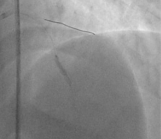
The patient underwent emergency coronary angiography, which revealed acute thrombotic ostial LAD (left anterior descending artery) occlusion and acute thrombotic ostial ramus intermedius (RI) near-occlusion. The right coronary artery (RCA) and the circumflex artery (LCx) did not present any significant lesions. (Figure 2) Therefore, we decided to perform LAD angioplasty. We intended to administer glyco-protein IIb/IIIa inhibitors, but none were available in our clinic. Two 0.014” angioplasty guidewires were passed into the distal LAD and RI and we performed thrombus aspiration, extracting a large coronary thrombus from the proximal segment of the LAD. (Fi-gure 3) This procedure was followed by the artery’s reperfusion and we noticed that the guidewire was actually a large diagonal branch (larger than the LAD) that provided septal perforating arteries and that the LAD was occluded in the distal segment (Figure 4). We implanted a 3.5x16mm everolimus eluting stent in the proximal LAD segment (Figure 5). Then, we passed the guidewire in the occluded LAD segment, intending to perform distal LAD angioplasty.
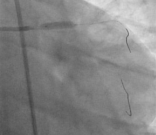
We performed thrombus aspiration once again, but the artery remained occluded. Then, we predilatated using a 1.5x15mm balloon, followed by a 2.25x20mm balloon (Figure 6), but we still could not obtain any coronary flow (TIMI 0 flow in the distal LAD). Taking into consideration the fact that the patient’s pain decreased, that the main septal perforating artery was before the occlusion site, and that the ECG monitor showed accelerated idioventricular rhythm, we decided that it would be safe to stop the procedure.
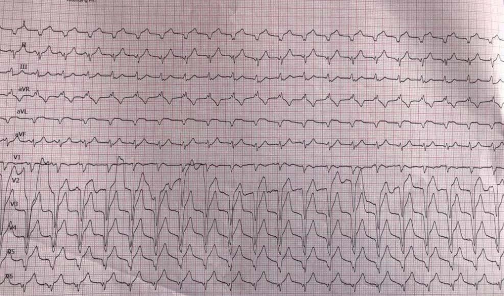
The patient was admitted in the coronary intensive care unit and was administered aspirin, ticagrelor, unfractionated heparin (aPTT adjusted dose), atorvastatin, enalapril, metoprolol and spironolactone. Significant reperfusion criteria were noticed: lack of chest pain, ST-segment elevation decrease, and recurrent accelerated idioventricular rhythm (Figure 7).
In order to check if we had to perform RI angioplasty before discharge, 8 days later, we performed another coronary angiography and, surprisingly, there was very few residual thrombi in the previously occluded LAD segment, and there was no more thrombus in the RI. We noticed TIMI 3 flow in all coronary arteries (Figure 8). Echocardiography performed before discharge showed a slight increase on the left ventricular systolic function, with an ejection fraction of 42%. The patient was discharged without any symptoms, and was scheduled for another medical examination within 6 weeks.

Figure 1. Initial emergency room ECG: normal sinus rhythm, ST-segment elevation in DI, aVL, V2-V4, and ST-segment depression in DIII and aVF

Figure 2. Coronary angiogram – left coronary artery (CAUD 40°): ostial LAD thrombotic occlusion (blue arrow), proximal RI thrombus (red arrow).

Figure 3. Coronary thrombus extracted from the proximal segment of the LAD.

Figure 4. Coronary angiography – left coronary artery (RAO 10°, CRAN 30°): distal LAD occlusion.

Figure 5. Proximal LAD stent implantation (3.5x16mm everolimus eluting stent); 0.014” guidewires placed in the distal diagonal branch and distal RI

Figure 6. Inflated 2.25x20mm balloon in the distal LAD occlusion area.
DISCUSSION
In the last two decades mortality from cardiovascular disease associated mortality has decreased in western countries. In older populations, the incidence of acute coronary syndromes has decreased, but this decline has not been noticed in younger people (especially men). Traditional risk factors are less common in young patients, and myocardial infarction is more frequently caused by spontaneous coronary artery dissection, vasospasm, and drug use. Coronary artery anomalies can also be involved, and these are easily identified during coronary angiography5. How
ever, plaque rupture still accounts for 60% to 65% of all myocardial infarction cases in young individuals6. Thrombophilia should also be considered in patients with myocardial infarction caused by large coronary thrombus7,8, and we believe that our patient should also be tested for these conditions and if a particular high-risk thrombophilia syndrome will be identified, oral anticoagulation should be considered.
One of the main particular features regarding this case is the large quantity of intracoronary thrombi. We decided to perform manual thrombus aspiration although the current guidelines based on recent trials do not support the routine use of this procedure4,9,10. However, we managed to extract a large, but we de-cided not to repeat the same procedure for the RI lesion, taking into consideration the fact that this artery was not occluded and several complications could occur. Another particular aspect regarding this case is the fact that we tried to perform manual thrombus aspiration for the distant LAD lesion, but without any success, even if this occlusion was likely caused by a distal embolization from the proximal thrombus. It is known that manual thrombus aspiration fails in over 30% of all cases11, but we also tried catheter balloon dilatation and failed. In relation to the high thrombus burden in our patient, a surprising result was the excellent response to unfractionated heparin, a drug with a high pharmacokinetic variability between patients12.
Regarding our patient’s coronary anatomy, a large diagonal branch with septal perforating arteries was found. The diameter of this diagonal artery was larger than that of the LAD (distal to the bifurcation). However, this is not a case of dual LAD, since there is no short LAD branch and the origin of the large diagonal artery was not that proximal13. Fortunately, the distal LAD occlusion occurred after the origin of two main septal perforating arteries (as seen in Fig. 4), therefore limiting the left ventricular septum ischemia after the initial angioplasty. Reperfusion of the occluded LAD segment occurred up to 8 days after the on-set of the infarction, and a further echocardiographic assessment of the patient is required to see if further improvement of the left ventricular systolic function will be identified.

Figure 7. Post-angioplasty ECG: accelerated idioventricular rhythm, HR= 110/min.

Figure 8. Coronary angiography – left coronary artery (RAO 10°, CRAN 30°): TIMI 3 flow in all coronary arteries. Small residual thrombus in the distal LAD.
CONCLUSION
Myocardial infarction can occur in young patients, even without many traditional risk factors. It is a po-tential life-threatening condition that requires emer-gency treatment. According to current guidelines, emergency PCI is the treatment of choice and manual thrombus aspiration can still be used in certain situa-tions, improving the patient’s outcome. The reported case outlines the role of PCI associated unfractionated heparin treatment and its continuous administration several days after the procedure.
Conflict of interest: none declared.
References
1. Entezarjou A, mohammad MA, Andell P, Koul S. Culprit vessel: im-pact on short-term and long-term prognosis in patients with ST-elevation myocardial infarction. Open Heart. 2018; 5: e000852.
2. Kobayashi N, Maehara A. Left anterior descending artery wrapping around the left ventricular apex predicts additional risk of future events after anterior myocardial infarction. Anatol J Cardiol. 2019; 21(5): 259-260.
3. Karha J, Murphy SA, Kirtane AJ, de Lemos JA, Aroesty JM, Cannon CP, Antman EM, Braunwald E, Gibson CM, TIMI Study Group. Evalu-ation of the association of proximal coronary culprit artery lesion location with clinical outcomes in acute myocardial infarction. The American Journal of Cardiology. 2003; 92(8): 913-918.
4. Ibanez B, James S, Agewall S, Antunes MJ, Bucciarelli-Ducci C, Bueno H, Caforio ALP, Crea F, Goudevenos JA, Halvorsen S, Hindricks G, Kastrati A, Lenzen MJ, Prescott E, Roffi M, Valgimigli M, Vaenhorst C, Vranckx P, Widimsky P. 2017 ESC Guidelines for the manage-ment of acute myocardial infarction in patients presenting with ST-segment elevation. European Heart Journal. 2018; 39: 119-177.
5. Ţieranu EN, Donoiu I, Istrătoaie O, Găman AE, Ţieranu ML, Ţieranu CG, Gheonea DI, Ciurea T. Rare case of single coronary artery in a patient with liver cirrhosis. Rom J Morphol Embryol. 2017; 58(4): 1505-1508.
6. Gulati R, Behfar A, Narula J, Kanwar A, Leman A, Cooper L, Singh M. Acute Myocardial Infarction in Young Individuals. Mayo Clin Proc. 2020; 95(1): 136-156.
7. Tahir F, Maijd Z, Khan S. Acute Myocardial Infarction as an Initial Presentation of Protein C and Protein S Deficiency Followed by Di-lated Cardiomyopathy in a Young Male. Cureus. 2019; 11(4): e4492.
8. Stepien K, Nowak K, Wypasek E, Zalewski J, Undas A. High preva-lence of inherited thrombophilia and antiphospholipid syndrome in myocardial infarction with non-obstructive coronary arteries: Com-parison with cryptogenic stroke. International Journal of Cardiology. 2019; 290: 1-6.
9. Frobert O, Lagerqvist B, Olivecrona G, Omerovic E, Gudnason T, Maeng M, Aasa M, Angeras O, Calais F, Danielewicz M, Erlinge D, Hellsten L, Jensen U, Johansson AC, Karegren A, Nilsson J, Robert-son L, Sandhall L, Sjogren I, Ostlund O, Harnek J, James SK. Throm-bus Aspiration during ST-Segment Elevation Myocardial Infarction. N Engl J Med. 2013; 369: 1587-1597.
10. Jolly SS, Cairns JA, Lavi S, Cantor WJ, Bernat I, Cheema AN, Moreno R, Kedev S, Stankovic G, Rao SV, Meeks B, Chowdhary S, Gao P, Sib-bald M, Velianou JL, Mehta SR, Tsang M, Sheth T, Dzavik V, TOTAL Investigators. Thrombus Aspiration in Patients With High Thrombus Burden in the TOTAL Trial. J Am Coll Cardiol. 2018; 72(14): 1589-1596.
11. Rao DS, Barik R. Thrombus aspiration catheter is a Dottering bal-loon. Indian Heart J. 2016; 68(4): 525-526.
12. Onwordi ENC, Gamal A, Zaman A. Anticoagulant Therapy for Acute Coronary Syndromes. Interv Cardiol. 2018; 13(2): 87-92.
13. Dhanse S, Kareem H, Rao MS, Devasia T. Dual LAD system- A case report and lessons learnt from past nomenclature system. IHJ Car-diovascular Case Reports. 2018; 2(1): 4-7.
 This work is licensed under a